
Журнал «Здоровье ребенка» Том 20, №4, 2025
Вернуться к номеру
Аналіз факторів ризику аритмії, аортальної та трикуспідальної регургітації в дітей із дефектом міжшлуночкової перегородки після транскатетерного закриття
Авторы: Alif Mutahhar, Mahrus A. Rahman
Faculty of Medicine, Universitas Airlangga, Surabaya, Indonesia
Department of Child Health, Dr. Soetomo General Academic Hospital, Surabaya, Indonesia
Рубрики: Педиатрия/Неонатология
Разделы: Клинические исследования
Версия для печати
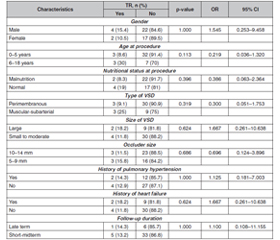
Актуальність. Дефект міжшлуночкової перегородки (ДМШП) — поширена вроджена вада серця в дітей, що зустрічається з частотою 5,27 випадку на 1000 живонароджених. Транскатетерне закриття ДМШП є потенційно ефективною альтернативою хірургічному втручанню завдяки меншій інвазивності, однак може супроводжуватися ускладненнями, зокрема аритмією, аортальною та трикуспідальною регургітацією. Мета: визначити поширеність і фактори ризику аритмії, аортальної та трикуспідальної регургітації після транскатетерного закриття ДМШП. Матеріали та методи. Поперечне дослідження було проведено в медичному закладі Dr. Soetomo Hospital з січня 2017 року до листопада 2024 року серед дітей із ДМШП після транскатетерного закриття. Для виявлення потенційних факторів ризику виконано двовимірний аналіз із використанням програмного забезпечення Statistical Package for the Social Sciences версії 22.0. Результати. У дослідженні взяли участь 45 дітей, переважно з нормальним харчовим статусом. Найпоширенішими типами ДМШП були перимембранозний і дефект середнього розміру. Більшість учасників не мали в анамнезі легеневої гіпертензії або серцевої недостатності. Післяпроцедурними ускладненнями були аритмія (17,8 %), трикуспідальна (13,3 %) та аортальна регургітація (11,1 %). Не виявлено вірогідних факторів, що збільшують ризик ускладнень після втручання. Висновки. Аритмія є найпоширенішим ускладненням транскатетерного закриття ДМШП, на яке не впливають соціально-демографічні й клінічні характеристики пацієнтів як потенційні фактори ризику. Моніторинг і подальше спостереження мають вирішальне значення щодо раннього виявлення можливих проблем і їхнього своєчасного усунення.
Background. Ventricular septal defect (VSD) is a prevalent congenital heart disease in children, occurring at a rate of 5.27 per 1000 live births. Transcatheter VSD closure is a potentially effective alternative to surgery, considering its less invasive nature, but may cause arrhythmia, aortic and tricuspid regurgitation as possible complications. This study aims to determine the prevalence and the risk factors for arrhythmia, aortic, and tricuspid regurgitation following transcatheter VSD closure. Materials and methods. A cross-sectional study was conducted at the Dr. Soetomo Hospital from January 2017 to November 2024 in children with VSD following transcatheter closure. Bivariate analysis was done to determine the potential risk factors using Statistical Package for the Social Sciences software version 22.0. Results. The study involved 45 participants, mainly with normal nutritional status. The most common types of VSD were perimembranous and moderate-sized. Most participants didn’t have a history of pulmonary hypertension or heart failure. Post-procedure complications were arrhythmia (17.8 %), tricuspid regurgitation (13.3 %), and aortic regurgitation (11.1 %). No significant factors were found to increase the risk of complications following the intervention. Conclusions. Arrhythmia is the most common complication after VSD transcatheter closure, not influenced by socio-demographic and clinical characteristics as potential risk factors. Monitoring and follow-up care are crucial to identify any potential issues early, allowing for timely intervention.
аритмія; дефект міжшлуночкової перегородки; транскатетерне закриття; аортальна регургітація; трикуспідальна регургітація
arrhythmia; ventricular septal defect; transcatheter closure; aortic regurgitation; tricuspid regurgitation
Для ознакомления с полным содержанием статьи необходимо оформить подписку на журнал.
- Fang G-H, еt al. The comparison of perventricular device closure with transcatheter device closure and the surgical repair via median sternotomy for perimembranous ventricular septal defect. Ann Thorac Cardiovasc Surg. 2018;24(6):308-314. doi: 10.5761/atcs.oa.18-00085.
- Li X, et al. Prediction of spontaneous closure of isolated ventricular septal defects in utero and postnatal life. BMC Pediatr. 2016;16(1):207. doi: 10.1186/s12887-016-0735-2.
- Penny DJ, Vick GW. Ventricular septal defect. Lancet. 2011;377(9771):1103-1112. doi: 10.1016/S0140-6736(10)61339-6.
- Lindinger A, Schwedler G, Hense H-W. Prevalence of congenital heart defects in newborns in Germany: results of the first registration year of the PAN study (July 2006 to June 2007). Klin Padiatr. 2010;222(5):321-326. doi: 10.1055/s-0030-1254155.
- Bergmann M, Germann CP, Nordmeyer J, Peters B, Berger F, Schubert S. Short- and long-term outcome after interventional VSD closure: a single-center experience in pediatric and adult patients. Pediatr Cardiol. 2021;42(1):78-88. doi: 10.1007/s00246-020-02456-2.
- Zhang K, et al. Aortic regurgitation requiring unplanned surgery following transcatheter closure of ventricular septal defect in children: incidence and risk factors. Cardiology. 2023;148(1):62-71. doi: 10.1159/000528115.
- Zheng Q, et al. A comparative study: early results and complications of percutaneous and surgical closure of ventricular septal defect. Cardiology. 2009;114(4):238-243. doi: 10.1159/000232405.
- Li H, et al. Short- and medium-term follow-up of transcatheter closure of perimembranous ventricular septal defects. BMC Cardiovasc Disord. 2019;19(1):222. doi: 10.1186/s12872-019-1188-y.
- Kanaan M, Ewert P, Berger F, Assa S, Schubert S. Follow-up of patients with interventional closure of ventricular septal defects with Amplatzer duct occluder II. Pediatr Cardiol. 2015;36(2):379-385. doi: 10.1007/s00246-014-1017-0.
- Yang P, et al. Unplanned surgery after transcatheter closure of ventricular septal defect in children: causes and risk factors. Front Pediatr. 2021;9:772138. doi: 10.3389/fped.2021.772138.
- Elmarsafawy H, Hafez M, Alsawah GA, Bakr A, Rakha S. Long-term outcomes of percutaneous closure of ventricular septal defects in children using different devices: a single centre experience from Egypt. BMC Pediatr. 2023;23(1):381. doi: 10.1186/s12887-023-04194-9.
- Sarkislali K, Kalangos A. Late tricuspid regurgitation after percutaneous transcatheter closure of ventricular septal defect: an educational presentation. Braz J Cardiovasc Surg. 2021;36(2). doi: 10.21470/1678-9741-2020-0172.
- Li Y, Hua Y, Fang J, Wan C, Wang C, Zhou K. Identification of risk factors for arrhythmia post transcatheter closure of perimembranous ventricular septal defect. J Invasive Cardiol. 2015;27(8):E158-E166.
- Ghosh S, Sridhar A, Sivaprakasam M. Complete heart block following transcatheter closure of perimembranous VSD using Amplatzer duct occluder II. Catheter Cardiovasc Interv. 2018;92(5):921-924. doi: 10.1002/ccd.27177.
- Mandal KD, Su D, Pang Y. Long-term outcome of transcatheter device closure of perimembranous ventricular septal defects. Front Pediatr. 2018;6:128. doi: 10.3389/fped.2018.00128.
- Salsabila N, Hidayat T, Lefi A. Characteristics of patients with ventricular septal defect that had been closed with transcatheter in Dr. Soetomo General Hospital from January 2019 to December 2021. Int J Res Publ. 2022;114(1). doi: 10.47119/ijrp10011411220224210.
- Klingberg E, Sveälv BG, Täng MS, Bech-Hanssen O, Forsblad-d’Elia H, Bergfeldt L. Aortic regurgitation is common in ankylosing spondylitis: time for routine echocardiography evaluation? Am J Med. 2015;128(11):1244-1250.e1. doi: 10.1016/j.amjmed.2015.04.032.
- Han Y, Li H, Zhu H, Sun G, Yin Q, Gu C. Aortic regurgitation after closure of ventricular septal defect by transcatheter device: the long-term complication. Cardiol Young. 2020;30(4):577-579. doi: 10.1017/S1047951120000414.
- Hamdar H, Ghrayeb H, Fakhry B, Chammas E, Chehab G. Treatment of ventricular septal defect in children: who, when, and how? A 20-years Lebanese multicentric retrospective study. World J Adv Res Rev. 2022;14(1):324-335. doi: 10.30574/wjarr.2022.14.1.0333.
- Sarmila B, Iskandar B, Daud D. Diagnostic value of electrocardiography for ventricular septal defect. Paediatr Indones. 2019;59(2):87-91. doi: 10.14238/pi59.2.2019.87-91.
- Lin L, et al. Risk factors for atrioventricular block after occlusion for perimembranous ventricular septal defect. Heart Rhythm. 2022;19(3):389-396. doi: 10.1016/j.hrthm.2021.11.027.
- Yulianti AC, Murni IK, Noormanto N, Nugroho S. Predictors of transcatheter closure cancellation in children with ventricular septal defect. Paediatr Indones. 2021;61(6):311-316. doi: 10.14238/pi61.6.2021.311-6.
- Lei Y-Q, et al. Influence of percutaneous catheter intervention for congenital perimembranous ventricular septal defects in children on the cardiac conduction system and associated risk factors: a meta-ana–lysis. J Cardiothorac Surg. 2022;17(1):19. doi: 10.1186/s13019-022-01751-8.
- Devendran V, Koneti NR, Jesudian V. Transcatheter closure of multiple perimembranous ventricular septal defects with septal aneurysm using two overlapping Amplatzer duct occluders II. Pediatr Cardiol. 2013;34(8):1963-1965. doi: 10.1007/s00246-012-0509-z.
- Lin Z-W, et al. The short and midterm follow-up of transthoracic device closure of perimembranous ventricular septal defect in adults. Heart Surg Forum. 2018;21(4):E242-E246. doi: 10.1532/hsf.1932.
- Obongonyinge B, Namuyonga J, Tumwebaze H, Aliku T, Lwabi P, Lubega S. Congenitally corrected transposition of great arte–ries: a case series of five unoperated African children. J Congenit Cardiol. 2020;4(1):8. doi: 10.1186/s40949-020-00038-8.
- Yang L, Tai B, Khin LW, Quek SC. A systematic review on the efficacy and safety of transcatheter device closure of ventricular septal defects (VSD). J Interv Cardiol. 2014;27(3):260-272. doi: 10.1111/joic.12121.
- Kozlik-Feldmann R, et al. Long-term outcome of perimembranous VSD closure using the Nit-Occlud® Lê VSD coil system. Clin Res Cardiol. 2021;110(3):382-390. doi: 10.1007/s00392-020-01750-6.
- Yildiz K, et al. Safety and efficacy of Amplatzer duct occluder II and Konar-MF VSD occluder in the closure of perimembranous ventricular septal defects in children weighing less than 10 kg. Front Cardiovasc Med. 2023;10. doi: 10.3389/fcvm.2023.1255808.
- Huang JS, Sun KP, Huang ST, Chen Q, Chen LW, Kuo YR. A meta-analysis of perventricular device closure of doubly committed subarterial ventricular septal defects. J Cardiothorac Surg. 2020;15(1):28. doi: 10.1186/s13019-020-01062-0.
- Polat TB, Türkmen E. Transcatheter closure of ventricular septal defects using the Amplatzer duct occluder II device: a single-center experience. Adv Interv Cardiol. 2016;4:340-347. doi: 10.5114/aic.2016.63635.
- Nguyen HL, et al. Nit-Occlud Lê VSD coil versus duct occluders for percutaneous perimembranous ventricular septal defect closure. Congenit Heart Dis. 2018;13(4):584-593. doi: 10.1111/chd.12613.
- Elsharkawy IM, Elwakeel AM, Elwakeel MM, Lamloom AH. Pulmonary artery venting in ventricular septal defects with pulmonary hypertension compared to ordinary routes of left ventricular venting. Egypt J Hosp Med. 2022;89(1):4323-4326. doi: 10.21608/ejhm.2022.256619.
- Hegazy YY, Koriem M, Keshk-Hegazy NS, Sodian R. Management of a residual VSD 60 years after one of the first operations worldwide. Thorac Cardiovasc Surg Rep. 2021;10(1):e22-e24. doi: 10.1055/s-0040-1722734.
- Aryal M, Timala R. Late aortic insufficiency after ventricular device closure: a case report. Clin Cardiol Cardiovasc Interv. 2022;5(6):1-3. doi: 10.31579/2641-0419/263.
- Alshahrani D, et al. Transfemoral perimembranous ventricular septal defect device closure in infants weighing < 10 kg. Pediatr Cardiol. 2023;44(5):1176-1182. doi: 10.1007/s00246-023-03100-5.
- Lu J, Lian X, Wen P, Liu Y. Case report: recovery of long-term delayed complete atrioventricular block after minimally invasive transthoracic closure of ventricular septal defect. Front Cardiovasc Med. 2023;10. doi: 10.3389/fcvm.2023.1226139.
- Yang J, et al. Transcatheter versus surgical closure of perimembranous ventricular septal defects in children. J Am Coll Cardiol. 2014;63(12):1159-1168. doi: 10.1016/j.jacc.2014.01.008.