Архив офтальмологии Украины Том 13, №1, 2025
Вернуться к номеру
Ефективність різних методів ексимерлазерних втручань у комплексному лікуванні кератоконуса
Авторы: Афанасьєв І.В.
ТОВ «Ексімер Одеса», м. Одеса, Україна
Рубрики: Офтальмология
Разделы: Клинические исследования
Версия для печати
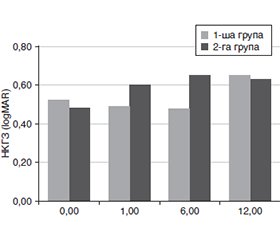
Актуальність. Кератоконус — дегенеративне захворювання, що призводить до погіршення якості зору, зниження гостроти зору, неможливості використання стандартних оптичних методів корекції аметропії та інколи — до сліпоти. Поширеність цього захворювання коливається від 50 до 265 на 100 000 населення. Сучасне лікування кератоконуса полягає не тільки в зупинці ектатичного процесу, але й у корекції аномалій рефракції. Тому нам видається актуальним дослідження ефективності та результатів лікування методиками крослінкінгу (CXL), поєднаними з ад’ювантними рефракційними процедурами, та їхнього впливу на корекцію аметропії. Мета: дослідити ефективність різних методів ексимерлазерних втручань у комплексному лікуванні кератоконуса при терміні спостереження 1 рік. Матеріали та методи. У дослідження було включено 58 пацієнтів (64 ока). Серед них було 40 чоловіків (69 %) і 18 жінок (31 %) віком від 20 до 40 років, які були розподілені на 2 групи залежно від методу втручання. Пацієнтам 1-ї групи виконано фоторефракційну кератектомію під контролем топографії з конвенційним (дрезденським) крослінкінгом (TG PRK з CXL). Пацієнтам 2-ї групи виконано трансепітеліальну фототерапевтичну кератектомію під контролем топографії (без корекції рефракційного компонента) з конвенційним (дрезденським) крослінкінгом (TG t-PTK з CXL). Результати. TG PRK з CXL і TG t-PTK з CXL були ефективними методиками для стабілізації рогівки й поліпшення гостроти зору в пацієнтів з кератоконусом 1–3-ї стадії. TG PRK з CXL мала перевагу щодо стабільного поліпшення максимально коригованої гостроти зору (МКГЗ), але асоціювався з вищою частотою ускладнень, зокрема з помутнінням рогівки й тривалішою епітелізацією. Натомість TG t-PTK з CXL характеризувалася меншою кількістю ускладнень і швидшим відновленням епітелію, хоча мала менш сприятливий вплив на некориговану гостроту зору (НКГЗ) у довгостроковій перспективі. Висновки. У результаті проведених нами досліджень встановлено, що TG PRK з CXL і TG t-PTK з CXL були достатньо ефективними в стабілізації рогівки й поліпшенні гостроти зору в пацієнтів з кератоконусом. Обидві групи показували поліпшення МКГЗ, але погіршення НКГЗ через 12 місяців без статистично значущої різниці між групами. Для обох технологій характерні ускладнення, однак їх кількість була вищою в 1-й групі спостереження без значущих міжгрупових відмінностей (p > 0,05), а помутніння рогівки через рік спостерігалося у 8 разів частіше в 1-й групі (23,5 % проти 3 %, p = 0,029), що, можливо, асоційовано з тривалішою епітелізацією (5,41 ± 0,45 доби для 1-ї групи і 4,70 ± 0,25 доби для 2-ї групи, p = 0,048). На наш погляд, перспективами подальших досліджень є вивчення й аналіз нових факторів ризику, етіологічних і генетичних чинників, які можуть оптимізувати вибір методики для конкретного пацієнта. Подальші дослідження мають бути спрямовані на вивчення оптимальних параметрів процедур (наприклад, глибини й об’єму абляції, дози ультрафіолету в крослінкінгу), а також на рефракційні зміни.
Background. Keratoconus is a degenerative disease that results in progressive vision deterioration, reduced visual acuity, inability to use standard optical methods for correcting ametropia, and, in some cases, blindness. The prevalence of this condition ranges from 50 to 265 cases per 100,000 individuals. Modern keratoconus treatment aims not only to halt the ectasia but also to correct refractive errors. Therefore, evaluating the effectiveness and outcomes of crosslinking (CXL) techniques combined with adjuvant refractive procedures and their impact on ametropia correction appears highly relevant. The purpose was to investigate the effectiveness of various methods for excimer laser interventions in the comprehensive treatment of keratoconus with a follow-up period of 1 year. Materials and methods. The study included 58 patients (64 eyes), comprising 40 men (69 %) and 18 women (31 %) aged 20 to 40 years. Participants were divided into two groups based on the intervention method. Group 1 underwent topography-guided photorefractive keratectomy combined with conventional (Dresden) crosslinking (TG PRK with CXL). Group 2 included patients who underwent topography-guided transepithelial phototherapeutic keratectomy (without correction of the refractive component) with conventional (Dresden) crosslinking (TG t-PTK with CXL). Results. TG PRK with CXL and TG t-PTK with CXL were effective methods for stabilizing the cornea and improving visual acuity in patients with keratoconus stages 1–3. TG PRK with CXL demonstrated superior sustained improvement in best-corrected visual acuity but was associated with a higher incidence of complications, including corneal opacity and prolonged epithelialization. In contrast, TG t-PTK with CXL resulted in fewer complications and faster epithelial recovery, though it had a less favorable long-term effect on uncorrected visual acuity. Conclusions. Our studies demonstrated that TG PRK with CXL and TG t-PTK with CXL were sufficiently effective in stabilizing the cornea and improving visual acuity in patients with keratoconus. Both groups exhibited an improvement in best-corrected visual acuity but a decline in uncorrected visual acuity at 12 months, with no statistically significant difference between the groups. Both techniques were associated with complications, though their frequency was higher in group 1; however, intergroup differences were not statistically significant (p > 0.05). Notably, corneal opacity after one year was eight times more frequent in group 1 (23.5 vs. 3 %, p = 0.029), possibly due to prolonged epithelialization (5.41 ± 0.45 days in group 1 vs. 4.70 ± 0.25 days in group 2, p = 0.048). We see potential for further research in exploring and analyzing new risk factors, as well as etiological and genetic factors, to optimize the selection of techniques for individual patients. Future studies should focus on determining the optimal procedural parameters (e.g., depth and volume of ablation, ultraviolet light dosage in CXL) and evaluating refractive changes.
кератоконус; фоторефракційна кератектомія під контролем топографії; фототерапевтична кератектомія під контролем топографії; комбіноване лікування
keratoconus; topography-guided photorefractive keratectomy; topography-guided phototherapeutic keratectomy; combined treatment
Для ознакомления с полным содержанием статьи необходимо оформить подписку на журнал.
- Li Х, Rabinowitz YS, Rasheed K, Yang H. Longitudinal study of the normal eyes in unilateral keratoconus patients. Ophthalmology. 2004;111:440-446. doi: 10.1016/j.ophtha.2003.06.020.
- Nichols JJ, Steger-May K, Edrington TB, Zadnik K. The relation between disease asymmetry and severity in keratoconus. Br J Ophthalmol. 2004;88:788-791. doi: 10.1136/bjo.2003.034520.
- Burns DM, Johnston FM, Frazer DG, Patterson C, Jackson AJ. Keratoconus: an analysis of corneal asymmetry. Br J Ophthalmol. 2004;88:1252-1255. doi: 10.1136/bjo.2003.033670.
- Jones-Jordan LA, Walline JJ, Sinnott LT, Kymes SM, Zad–nik К. Asymmetry in keratoconus and vision-related quality of life. Cornea. 2013;32:267-272. doi: 10.1097/ICO.0b013e31825697c4.
- Chopra І, Jain AK. Between eye asymmetry in keratoconus in an Indian population. Сlin Exp Optom. 2005;88:146-152. doi: 10.1111/j.1444-0938.2005.tb06687.x.
- Zadnik K, Steger-May K, Fink BA, Joslin CE, Nichols JJ, Rosenstiel CE et al. Between-eye asymmetry in keratoconus. Cornea. 2002;21:671-679. doi: 10.1097/00003226-200210000-00008.
- Santodomingo-Rubido J, Carracedo G, Suzaki A, Villa-Collar C, Vincent SJ, Wolffsohn JS. Keratoconus: An updated review. Cont Lens Anterior Eye. 2022 Jun;45(3):101559. doi: 10.1016/j.clae.2021.101559. Epub 2022 Jan 4. PMID: 34991971.
- Lucas SEM, Burdon KP. Genetic and Environmental Risk Factors for Keratoconus. Annu Rev Vis Sci. 2020;6:25-46.
- Toprak І, Vega А, Alió Del Barrio JL, Espla E, Cavas F, Alió JL. Diagnostic value of corneal epithelial and stromal thickness distribution profiles in forme fruste keratoconus and subclinical keratoconus. Cornea. 2021;40:61-72. doi: 10.1097/ICO.0000000000002435.
- O’Brart DP, Patel P, Lascaratos G, Wagh VK, Tam C, Lee J, O’Brart NA. Corneal cross-linking to halt the progression of keratoconus and corneal ectasia: seven-year follow-up. Am J Ophthalmol. 2015;160(6):1154-1163. doi: 10.1016/j.ajo.2015.08.023.
- Raiskup F, Theuring A, Pillunat LE, Spoerl E. Corneal collagen crosslinking with riboflavin and ultraviolet-A light in progressive keratoconus: ten-year results. J Cataract Refract Surg. 2015;41(1):41-46. doi: 10.1016/j.jcrs.2014.09.033.
- Grentzelos MA, Kounis GA, Diakonis VF, Siganos CS, Tsilimbaris MK, Pallikaris IG, Kymionis GD. Combined transepithelial phototherapeutic keratectomy and conventional photorefractive keratectomy followed simultaneously by corneal crosslinking for keratoconus: Cretan protocol plus. J Cataract Refract Surg. 2017;43:1257-1262. doi: 10.1016/j.jcrs.2017.06.047.
- Zhu W, Han Y, Cui C, Xu W, Wang X, Dou X, Xu L, Xu Y, Mu G. Corneal collagen crosslinking combined with phototherapeutic keratectomy and photorefractive keratectomy for corneal ectasia after laser in situ keratomileusis. Ophthalmic Res. 2018;59:135-141. doi: 10.1159/000480242.
- Wollensak G, Spoerl E, Seiler T. Riboflavin/ultraviolet-A-–indu–ced collagen crosslinking for the treatment of keratoconus. Am J Ophthal–mol. 2003;135(5):620-627. doi: 10.1016/S0002-9394(02)02220-1.
- Wisse RPL, Kuiper JJW, Gans R, Imhof S, Radstake TRDJ, Van Der Lelij A. Cytokine expression in keratoconus and its corneal microenvironment: A systematic review. Ocul Surf. 2015;13:272-283. doi: 10.1016/j.jtos.2015.04.006.
- Jun AS, Cope L, Speck С, Feng Х, Lee S, Meng Н, et al. Subnormal cytokine profile in the tear fluid of keratoconus patients. PLOS One. 2011;6:е16437. doi: 10.1371/journal.pone.0016437.
- Balasubramanian SA, Mohan S, Pye DC, Willcox MDP. Proteases, proteolysis and inflammatory molecules in the tears of people with keratoconus. Acta Ophthalmol. 2012;90:303-309. doi: 10.1111/j.1755-3768.2011.02369.x.
- Galvis V, Sherwin T, Tello A, Merayo J, Barrera R, Acera A. Keratoconus: an inflammatory disorder? Eye. 2015;29:843-859. doi: 10.1038/eye.2015.63
- McMonnies CW. Inflammation and keratoconus. Optom Vis Sci. 2015;92:e35-e41. doi: 10.1097/OPX.0000000000000455.
- Amsler M. Kératocône classique et kératocône fruste; arguments unitaires. Ann Ocul (Paris). 1947;180:112. https://doi.org/10.1159/000300309.
- Ocular Surface Disease Index (OSDI). Administration and Scoring Manual Allergan, Inc., Irvine, CA. 2004.
- Fantes FE, Hanna KD, Waring GO, Pouliquen Y, Thomp–son KP, Savoldelli M. Wound healing after excimer laser keratomileusis (photorefractive keratectomy) in monkeys. Arch Ophthalmol. 1990;108: 665-675. doi: 10.1001/archopht.1990.01070070051034.
- Zhang H, Cantó-Cerdán M, Félix-Espinar B. Efficacy of Customized Photorefractive Keratectomy With Cross-Linking Versus Cross-Linking Alone in Progressive Keratoconus: A Systematic Review and Meta-Analysis. American Journal of Ophthalmology. 2025;274:9-23.
- Iqbal M, Elmassry A, Tawfik A, Elgharieb ME, El Deen Al Nahrawy OM, Soliman AH et al. Evaluation of the Effectiveness of Cross-Linking Combined With Photorefractive Keratectomy for Treatment of Keratoconus. Cornea. 2018 Sep;37(9):1143-1150. doi: 10.1097/ICO.0000000000001663. PMID: 29952798; PMCID: PMC6092093.
- Koller T, Mrochen M, Seiler T. Complication and failure rates after corneal crosslinking. Journal of Cataract & Refractive Surgery. 2009;35(8):1358-1362. DOI: 10.1016/j.jcrs.2009.03.035.
- Kanellopoulos AJ. Combined Photorefractive Keratectomy and Corneal Cross-Linking for Keratoconus and Ectasia: The Athens Protocol Cornea. 2023 Oct 1;42(10):1199-1205. doi: 10.1097/ICO.0000000000003320. Epub 2023 Jul 10. PMID: 37669421; PMCID: PMC10476591.
- Netto MV, Mohan RR, Sinha S et al. Stromal haze, myofibroblasts, and surface irregularity after PRK. Exp Eye Res. 2006;82(5):788-797.
- Cho MY, Kanellopoulos AJ. Short and Long-term Complications of Combined Topography-guided Photorefractive Keratectomy and Riboflavin/Ultraviolet A Corneal Collagen Cross-linking (the Athens Protocol) in 412 Keratoconus Eyes. Invest Ophthalmol Vis Sci. 2011;52(14):5202.
- Natarajan R, Giridhar D. Corneal scarring after epithelium-off collagen cross-linking. Indian Journal of Ophthalmology. 2025;73(1):28-34. DOI: 10.4103/IJO.IJO_95_24.
- Li L, Zhang B, Hu Y, Xiong L, Wang Z. Comparison of safety and efficiency of corneal topography-guided photorefractive keratectomy and combined with crosslinking in myopic correction: An 18-month follow-up. Medicine (Baltimore). 2021 Jan 15;100(2):e23769. doi: 10.1097/MD.0000000000023769. PMID: 33466126; PMCID: PMC7808543.
- Ohana O, Kaiserman I, Domniz Y, Cohen E, Franco O, Sela T, Munzer G, Varssano D. Outcomes of simultaneous photorefractive keratectomy and collagen crosslinking. Can J Ophthalmol. 2018 Oct;53(5):523-528. doi: 10.1016/j.jcjo.2017.12.003. Epub 2018 Feb 13. PMID: 30340722.
- Torricelli AA, Santhanam A, Wu J, Singh V, Wilson SE. The corneal fibrosis response to epithelial-stromal injury. Exp Eye Res. 2016 Jan;142:110-8. doi: 10.1016/j.exer.2014.09.012. PMID: 26675407; PMCID: PMC4683352.
- Bonzano C, Cutolo CA, Musetti D, Di Mola I, Pizzorno C, Scotto R, Traverso CE. Delayed re-epithelialization after epithe–lium-off crosslinking: predictors and impact on keratoconus progression. Front Med (Lausanne). 2021 Oct 15;8:657993. doi: 10.3389/fmed.2021.657993. PMID: 34722556; PMCID: PMC8554242.
- Bonzano С, Musetti D, Scotto R, Sturloni М, Cutolo CA, Traverso CE. Delayed re-epithelialization after epithelium-off crosslinking: associated factors and impact on keratoconus progression. Invest Ophthalmol Vis Sci. 2019;60(9):349.
- Gaeckle HC. Early clinical outcomes and comparison between trans-PRK and PRK, regarding refractive outcome, wound healing, pain intensity and visual recovery time in a real-world setup. BMC Ophthalmol. 2021;21:181. https://doi.org/10.1186/s12886-021-01941-3.
- Labiris G, Giarmoukakis A, Larin R, et al. Impact of preope–rative dry eye disease on epithelial healing after corneal cross-lin–king. Journal of Refractive Surgery. 2016;32(5):326-331. DOI: 10.3928/1081597X-20160225-01.
- Kanellopoulos AJ. Comparison of sequential vs same-day simultaneous collagen cross-linking and topography-guided PRK for treatment of keratoconus. Journal of Refractive Surgery. 2007;23(8):801-806. DOI: 10.3928/1081597X-20070901-08.
- Kontadakis GA, Kankariya VP, Tsoulnaras K. Long-term complications of combined topography-guided photorefractive keratectomy and corneal cross-linking. Journal of Cataract & Refractive Surgery. 2016;42(8):1139-1145. DOI: 10.1016/j.jcrs.2016.06.023.
- Kymionis GD, Grentzelos MA, Kankariya VP, et al. Long-term results of combined transepithelial phototherapeutic keratectomy and corneal collagen crosslinking for keratoconus. Cornea. 2012;31(11):1275-1280. DOI: 10.1097/ICO.0b013e31823e2d86.
- Alessio G, L’abbate M, Sborgia C, La Tegola MG. Photo–refractive keratectomy followed by cross-linking versus cross-lin–king alone for management of progressive keratoconus. American Journal of Ophthalmology. 2013;155(2):420-426. DOI: 10.1016/j.ajo.2012.08.021.
- Kremer I, Levinger E, Levinger S. Combined phototherapeutic keratectomy and corneal collagen cross-linking for keratoconus: A review of visual outcomes. Eye & Contact Lens. 2016;42(5):298-303. DOI: 10.1097/ICL.0000000000000190.

