Інформація призначена тільки для фахівців сфери охорони здоров'я, осіб,
які мають вищу або середню спеціальну медичну освіту.

Підтвердіть, що Ви є фахівцем у сфері охорони здоров'я.

Oral and General Health Том 5, №4, 2024

Вернуться к номеру

Склеротичний дентин: фізіологічні та патологічні аспекти його формування

Авторы: Савичук А.О., Паливода І.І., Корнієнко Л.В., Трубка І.О., Савичук Н.О.
Національний університет охорони здоров’я України імені П.Л. Шупика, м. Київ, Україна

Рубрики: Стоматология

Разделы: Справочник специалиста

Версия для печати


Резюме

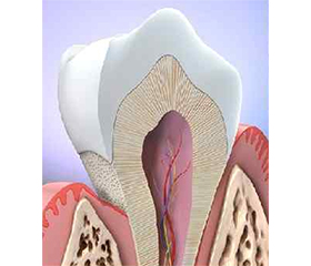
Актуальність. Склеротичний дентин формується під впливом вікових змін або у відповідь на патологічні фактори, такі як карієс, ерозія чи механічні пошкодження, включно з наслідками стоматологічного лікування. Ці зміни призводять до зниження еластичності й міцності дентину та його опору до утворення тріщин, що негативно впливає на адгезію реставраційних матеріалів. В умовах зростаючого попиту на довготривалий ефект лікування зубів у старших вікових групах особливо важливо розуміти вплив склеротичного дентину на результати лікування для подальшого вдосконалення протоколів надання стоматологічних послуг. Мета: дослідити структуру та механізми утворення склеротичного дентину, а також його вплив на механічні властивості зубів і ефективність адгезивних реставрацій; оцінити роль цих змін у контексті сучасних клінічних підходів до лікування. Висновки. Склеротичний дентин погіршує механічні властивості зуба, знижуючи його стійкість до навантажень і адгезію композитних матеріалів. Для оптимізації клінічних протоколів потрібні подальші дослідження, спрямовані на поліпшення ефективності реставрацій при лікуванні зубів з такими змінами.

Background. Sclerotic dentin forms as a result of ­aging or in response to pathological factors such as caries, erosion, or mechanical damage, including the effects of dental treatments. These changes reduce the elasticity and strength of dentin, promoting crack formation, which negatively impacts the adhesion of resto­rative materials. Given the growing demand for long-term treatment success in older patients, understanding the impact of sclerotic dentin on treatment outcomes is crucial for improving clinical protocols. The aim of this review was to investigate the structure and mechanisms of sclerotic dentin formation, as well as its impact on the mechanical properties of teeth and the effectiveness of adhesive restorations. The role of these changes in the context of mo­dern clinical approaches to treatment was evaluated. Conclusions. Sclerotic dentin deteriorates the mechanical properties of the tooth, reducing its resistance to loads and affecting the adhesion of composite materials. Further research is required to optimize clinical protocols and improve the effectiveness of restorations in teeth with these changes.


Ключевые слова

склеротичний дентин; старіння; адгезія; механічні властивості; реставрація

sclerotic dentin; aging; adhesion; mechanical properties; restoration


Для ознакомления с полным содержанием статьи необходимо оформить подписку на журнал.


Список литературы

1. Kinney JH, Nalla RK, Pople JA, Breunig TM, Ritchie RO. Age-related transparent root dentin: Mineral concentration, crystallite size, and mechanical properties. Biomaterials. 2005 Aug;26(16):3363-76. doi: 10.1016/j.biomaterials.2004.09.010.
2. Wilson EM, Green JR, Weismer G. A kinematic description of the temporal characteristics of jaw motion for early chewing: Preliminary findings. Journal of Speech, Language, and Hearing Research, 2012;55(2):626-638. 
3. Vasiliadis L, Darling AI, Levers BGH. The amount and distribution of sclerotic human root dentine. Archives of Oral Biology. 1983;28:645-649.
4. Arola D, Reprogel RK. Effects of aging on the mechanical behavior of human dentin. Biomaterials 2005;26:4051e61.
5. Bajaj D, Sundaram N, Nazari A, Arola D. Age, dehydration and fatigue crack growth in dentin. Biomaterials 2006;27:2507e17.
6. Murray PE, Stanley HR, Matthews JB, Sloan AJ, Smith AJ. Agerelated odontometric changes of human teeth. Oral Surg Oral Med Oral Pathol Oral Radiol Endod 2002;93:474-82.
7. Tay FR, Pashley DH. Resin bonding to cervical sclerotic dentin: A review. Journal of Dentistry. 2004;32(3):173-196.
8. Lin J, Bennani V, Aarts JM, Brunton P. Factors influencing success rate of ceramic veneers on endodontically treated anterior teeth: A systematic review. Journal of Prosthetic Dentistry. In Press. 2023.
9. World Population Prospects: The 2002 Revision. Volume II Sex and Age Distribution of Populations. United Nations New York. 2003.
10. Warren JJ, Cowen HJ, Watkins CM, Hand JS. Dental caries prevalence and dental care utilization among the very old. J Am Dent Assoc. 2000;131:1571-9.
11. Jones JA, Adelson R, Niesson LC, Gilbert GH. Issues in financing dental care for the elderly. J Public Health Dent. 1990;50:268-75.
12. Kagayama M, Sasano Y, Tssuchia M, Watanabe M, Mizoguchi I, Kamakura S, Motegi K. Confocal microscopy of Tomes’s granular layer in dog premolar teeth. Anat Embryol. 2000;201:131-137.
13. Wang RZ, Weiner S. Strain-structure relations in human teeth using Moiré fringes. J Biomech. 1998;31:135-141.
14. Linde A (Ed.). Secondary and reparative dentin formation. In Dentin and Dentinogenesis. CRC Press. 1984;107-120.
15. Wang R, Weiner S. Human root dentin: structural ani-sotropy and Vickers microhardness isotropy. Connect Tissue Res. 1998;39:269-279.
16. Schilke R, Lisson JA, Bauss O, Geurtsen W. Comparison of the number and diameter of dentinal tubules in human and bovine dentine by scanning electron microscopic investigation. Arch Oral Biol. 2000;45:355-361.
17. Goldberg M, Septier D, Oldberg A, Young MF, Ameye LG. Fibromodulin-deficient mice display impaired collagen fibrillogenesis in predentin as well as altered dentin mineralization and enamel formation. J Histochem Cytochem. 2006;54:525-537.
18. Höehl E. Beitrag zur histology der pulpa und des dentins. Archiv für Anatomie und Physiologie. 1896;32:31-54.
19. Goldberg M, Septier D. A comparative study of the transition between predentin and dentin using various preparative procedures in the rat. Europ J Oral Sciences. 1996;104:269-277.
20. Bergman G, Engfeldt B. Studies on mineralised dental tissues II. Acta Odontologica Scandinavica, 1955-1956;13:1-7.
21. Weber DF. Human dentine sclerosis: A microradiographic survey. Archives of Oral Biology. 1974;19(12):1135-1142. 
22. Furseth R. The structure of peripheral root dentine in young human premolars. Scandinavian Journal of Dental Research, 1974;82:557-561.
23. Thomas GJ, Whittaker DK, Embery G. A comparative study of translucent apical dentine in vital and non-vital human teeth. Archives of Oral Biology. 1994;39(1):29-34.
24. Yu C. An overview of the dental pulp: its functions and responses to injury. Australian Dental Journal, 2008;53(S1):S1-S5. 
25. Weine FS, He B, Waxman B, Smulson M. Cytotoxicity of endodontic materials. Journal of Endodontics. 1985;11(10):415-420. 
26. Goldberg M, Kulkarni AB, Young M, Boskey A. Dentin: structure, composition and mineralization. Frontiers in Bioscience. 2011;E3(2):711-735. doi: 10.2741/e281.
27. Kabartai F, Hoffmann T, Hannig C. The physiologic sclerotic dentin: A literature-based hypothesis. Medical Hypotheses. 2017;105:52-55.
28. Miles AE. Ageing in the teeth and oral tissues. In: Bourne GH, editor. Structural aspects of ageing. London: Pitman. 1961;353-79.
29. Vasiliadis L, Stavrianos C, Dagkalis P, Parisi K, Pantelidou O, Samara E. Root dentine translucency of human teeth: factors affecting formation and deposition. Res J Biol Sci 2011;6(2):82-8.
30. Brännström M, Garberoglio R. The dentinal tubules and the odontoblast processes. Acta Odontologica Scandinavica. 1972;30:291-311.
31. Gupta HS, Seto J, Wagermaier W, Zaslansky P, Boesecke P, Fratzl P. Cooperative deformation of mineral and collagen in bone at the nanoscale. Proceedings of the National Academy of Sciences. 2006;103(47):17741-17746. 
32. Bonucci E. Calcification in biological systems. Boca Raton: CRC Press; 1992.
33. Pashley DH, Walton RE, Slavkin HC. Histology and physio–logy of the dental pulp. In: Ingle JI, Bakland LK, editors. Endodontics, Band 1. Ontario: PMPHUSA. 2002;25-61.
34. Fink SL, Cookson BT. Apoptosis, pyroptosis, and necrosis: mechanistic description of dead and dying eukaryotic cells. Infect Immun 2005;73(4):1907-16.
35. Savill J, Fadok V, Henson P, Haslett C. Phagocyte recognition of cells undergoing apoptosis. Immunol Today. 1993;14(3):131-6.
36. Christoffersen J. Christoffersen MRLB-S-R. Kinetics of dissolution of calcium hydroxyapatite. V. The acidity constant for the hydrogen phosphate surface complex. J Crystal Growth. 1982;57:21-6 
37. Versiani MA, Souza E, De-Deus G, Leoni GB. Critical appraisal of studies on dentinal tubule penetration of endodontic sealers. Journal of Endodontics. 2020;46(8):1039-1046.

Вернуться к номеру