Архив офтальмологии Украины Том 11, №3, 2023
Вернуться к номеру
Дослідження впливу ригідності рогівки на показники внутрішньоочного тиску при різних видах тонометрії у пацієнтів з еметропічною та міопічною рефракцією
Авторы: Пінчук Є.А.
Національний медичний університет імені О.О. Богомольця, м. Київ, Україна
Рубрики: Офтальмология
Разделы: Клинические исследования
Версия для печати
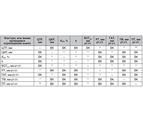
Актуальність. Визначення біомеханічних властивостей рогівки є актуальним науково-прикладним завданням сучасної клінічної офтальмології, оскільки рогівка є маркером змін біомеханічної поведінки ока. Низка досліджень свідчать, що біомеханіка рогівки змінюється у пацієнтів з міопією і залежить від ступеня міопії. Метою нашого дослідження було визначення впливу ригідності рогівки на показники внутрішньоочного тиску при різних видах тонометрії у пацієнтів з еметропією та міопією різного ступеня. Матеріали та методи. У дослідженні взяли участь 194 пацієнти (372 ока) з еметропією (60 очей) та міопічною рефракцією різних ступенів (312 очей). Середній вік пацієнтів становив 25 ± 2 року. Серед обстежених було 95 чоловіків (48,97 %) і 99 жінок (51,03 %). Визначення біомеханічних властивостей, коефіцієнта ригідності рогівки (KER) проводили з використанням формули та способу оцінки ригідності рогової оболонки ока (Сергієнко М.М., Шаргородська І.В., 2008). Для кожного ока проводили розрахунок внутрішньоочного тиску з урахуванням коефіцієнта ригідності рогівки — ВОТ(к) та поправочного коефіцієнта з урахуванням коефіцієнта ригідності рогівки — k. Результати. Аналіз результатів свідчив про вплив центральної товщини рогівки (ЦТР) на показники тонометрії при визначенні ВОТ на еметропічних очах методом тонометрії за Маклаковим (r = 0,532, р < 0,05), рикошетної тонометрії (r = 0,334, р < 0,05) і особливо пневмотонометрії (r = 0,611, р < 0,05). Найменший вплив ЦТР на показники тонометрії був визначений при апланаційній тонометрії Гольдмана (r = 0,186, р < 0,05). Результати свідчили про відсутність кореляції між коефіцієнтом ригідності рогівки на еметропічних очах та ЦТР, незалежність цього показника від рівня внутрішньоочного тиску, що підтверджувалося визначенням ВОТ різними методами, та вплив центральної кривизни рогівки (ЦКР) на KER. Було встановлено, що коефіцієнт ригідності рогівки міопічних очей залежав від ЦКР та корелював зі ступенем міопії. Водночас визначено відсутність кореляції KER міопічних очей з центральною товщиною рогівки. Значення ВОТ, отримані з використанням рикошетної тонометрії на міопічних очах, при міопії слабкого та середнього ступеня майже не відрізнялися від апланаційної тонометрії Гольдмана, а при міопії високого ступеня були значно нижчими (t = –2,63, P = 0,005). Висновки. У результаті проведеного дослідження встановлено, що визначення біомеханічних властивостей рогівки має велике значення для точного вимірювання внутрішньоочного тиску. Більш ефективною методикою є прижиттєве визначення коефіцієнта ригідності рогівки та врахування його як поправки при визначенні розрахункового ВОТ на еметропічних очах та очах з міопією різного ступеня.
Background. Determining the biomechanical properties of the cornea is an urgent applied scientific task of modern clinical ophthalmology, since the cornea is a marker of changes in the biomechanical behavior of the eye. A number of studies show that corneal biomechanics changes in patients with myopia and depends on its degree. The aim of our study was to determine the influence of corneal rigidity on intraocular pressure (IOP) indicators with different types of tonometry in patients with emmetropia and myopia of varying degrees. Materials and methods. One hundred and ninety-four patients (372 eyes) with emmetropia (60 eyes) and myopia (312 eyes) of varying degrees participated in the study. Their average age was 25 ± 2 years. Among the examined, there were 95 men (48.97 %) and 99 women (51.03 %). Determination of biomechanical properties, the coefficient of corneal rigidity (KER) was carried out using the formula and method for assessing the corneal rigidity (Sergienko M.M., Shargorodska I.V., 2008). For each eye, we calculated the intraocular pressure with the coefficient of corneal rigidity — IOP(k) and the correction factor taking into account the coefficient of corneal rigidity — k. Results. The analysis of the results demonstrated the influence of the central corneal thickness (CCT) on tonometry indicators when determining IOP in emmetropic eyes by the Maklakov method (r = 0.532, p < 0.05), ricochet tonometry (r = 0.334, p < 0.05) and especially pneumotonometry (r = 0.611, p < 0.05). The effect of CCT on tonometry was smallest in Goldman applanation tonometry (r = 0.186, p < 0.05). The results show the absence of a correlation between the coefficient of corneal rigidity in emmetropic eyes and the central corneal thickness, the independence of this indicator from the level of intraocular pressure, which was confirmed by the determination of IOP by various methods, and the effect of CCT on KER. It was found that the coefficient of corneal rigidity of myopic eyes depended on the CCT and correlated with the degree of myopia. At the same time, there was a lack of correlation between KER of myopic eyes and the central corneal thickness. The values of IOP obtained using ricochet tonometry on eyes with mild and moderate myopia almost did not differ from that of Goldmann applanation tonometry and were significantly lower with severe myopia (t = –2.63, p = 0.005). Conclusions. As a result of the conducted research, it was found that determining the biomechanical properties of the cornea is of great importance for accurate measurement of intraocular pressure. A more effective technique is the intravital calculation of the coefficient of corneal rigidity and its consideration as an adjustment when determining the estimated IOP in emmetropic eyes and eyes with varying degrees of myopia.
внутрішньоочний тиск; коефіцієнт ригідності рогівки; тонометрія; міопія; еметропія; рогівка
intraocular pressure; coefficient of corneal rigidity; tonometry; myopia; emmetropia; cornea
Для ознакомления с полным содержанием статьи необходимо оформить подписку на журнал.
- Pye D.C. A clinical method for estimating the modulus of elasticity of the human cornea in vivo. PLoS One. 2020. 15 (1). e0224824. doi: 10.1371/journal.pone.0224824.
- Dastiridou A.I. Ocular Rigidity and Intraocular Pressure. In: Pallikaris I., Tsilimbaris M.K., Dastiridou A.I. (eds). Ocular Rigidity, Biomechanics and Hydrodynamics of the Eye. Springer, Cham, 2021. https://doi.org/10.1007/978-3-030-64422-2_16.
- Girard M.J., Dupps W.J., Baskaram M., Scarcelli G., Yun S.H., Quigley H.A. et al. Translating ocular biomechanics into clinical practice: current state and future prospects. Curr. Eye Res. 2015. 40. 1-18. 10.3109/02713683.2014.914543.
- Dupps W.J., Roberts C.J. Corneal biomechanics: a decade later. J. Cataract. Ref. Surg. 2014. 40. 857.
- Luz A., Faria-Correia F., Salomao M.Q., Lopes B.T., Ambrosio R. Jr. Corneal biomechanics: Where are we? J. Curr. Ophthalmol. 2016. 97-8. 10.1016/j.joco.2016.07.004.
- Sergienko N.M., Shargorodska I.V. Determining corneal hysteresis and preexisting intraocular pressure. J. Cataract. Refract. Surg. 2009. 35. 2033-2034.
- Kotecha A. What biomechanical properties of the cornea are relevant to the clinician? Surv. Ophthalmol. 2007. 52 Suppl. 2. S109-14.
- Hon Y., Chen G.-Z., Lu S.-H., Lam D.C.C., Lam A.K.C. High myopes have lower normalised corneal tangent moduli (less ’stiff” corneas) than low myopes. Ophthalmic Physiol. Opt. 2016. 37. 42-50. 10.1111/opo.12335.
- Сергієнко М.М., Шаргородська І.В. Вивчення біомеханічних властивостей рогівки при міопії. Офтальмологічний журнал. 2011. 5(442). 24-26.
- Wan K., Cheung S.W., Wolffsohn J.S., Orr J.B., Cho P. Role of corneal biomechanical properties in predicting of speed of myopia progression in children wearing orthokeratology lenses or single-vision spectacles. BMJ Open. Ophth. 2018. 3. e000204 10.1136/bmjophth-2018-000204 eCollection 2018.
- Шаргородська І.В. Діагностика прогресуючої короткозорості. Проблеми екологічної та медичної генетики та клінічної імунології. Збірник наукових праць. 2012. 1 (109). 409-414.
- Ramm L., Herber R., Spoerl E., Pillunat L.E., Terai N. Measurement of corneal biomechanical properties in diabetes mellitus using the Ocular Response Analyzer and the Corvis ST. Cornea. 2019. 5. 595-99.
- Wu N., Chen Y., Yu X., Li M., Wen W., Sun X. Changes in corneal biomechanical properties after long-term topical prostaglandin therapy. PLoS One. 2016. 17. 11(5). e0155527. 10.1371/journal.pone.0155527 eCollection 2016.
- Zhao Y., Shen Y., Yan Z., Tian M., Zhao J., Zhou X. Relationship among stiffness, thickness, and biomechanical parameters measured by the Corvis ST, Pentacam and ORA in keratoconus. Front. Physiol. 2019. 13. 10. 740. 10.3389/fphys.2019.00740 eCollection 2019.
- Шаргородська І.В. Порівняльний аналіз виміру біомеханічних показників рогівки у пацієнтів з кератоконусом при використанні різних методів. Міжнародний науково-практичний журнал «Офтальмологія». 2016. 3 (05). 50-67.
- Belovay G.W., Goldberg I. The thick and thin of the central corneal thickness in glaucoma. Eye. 2018. 32. 915-923.
- Mahendradas P., Francis M., Vala R., Poornachandra B., Kawali A., Sheytty R. et al. Quantification of ocular biomechanics in ocular manifestations of systemic autoimmune diseases. Ocul. Immunol. Inflamm. 2018. August 7. 1-11. 10.1080/09273948.2018.1501491.
- Woo S.L., Kobayashi A.S., Schlegel W.A., Lawrence C. Nonlinear material properties of intact cornea and sclera. Exp. Eye Res. 1972. 14. 29-39. 10.1016/0014-4835(72)90139-x.
- Smolek M.K. Holographic interferometry of intact and radially excised human eye-bank corneas. J. Cataract. Refract. Surg. 1994. 20. 277-86. 10.1016/s0886-3350(13)80578-0.
- Tanter M., Touboul D., Gennisson J.L., Bercoff J., Fink M. High-resolution quantitative imaging of corneal elasticity using supersonic shear imaging. IEE Trans. Med. Imag. 2009. 28. 1881-93.
- Last J.A., Thomasy S.M., Croasdale C.R., Russell P., Murphy C.J. Compliance profile of the human cornea as measured by atomic force microscopy. Micron 43. 2012. 1293-98. 10.1016/j.micron.2012.02.014.
- Ford M.R., Sinha Roy A., Rollins A.M., Dupps W.J. Jr. Serial biomechanical comparison of edematous, normal, and collagen crosslinked human donor corneas using optical coherence tomography. J. Cataract. Refract. Surg. 2014. 40. 1041-7. 10.1016/j.jcrs.2014.03.017.
- Yun S.H., Chernyak D. Brilluoin microscopy: assessing ocular tissue biomechanics. Curr. Opin. Ophthalmol. 2018. 29. 299-305. 10.1097/ICU.0000000000000489.
- Lam A.K., Hon Y., Leung L.K., Lam D.C. Repeatability of a novel corneal indentation device for corneal biomechanical measurement. Ophthalmic Physiol. Opt. 2015. 35. 455-61. 10.1111/opo.12219.
- Shih P.-J., Cao H.-J., Hunag C.-J., Wang I.-J., Yen J.-Y. A corneal elastic dynamic model derived from Scheimpflug imaging technology. Ophthalmic Physiol. Opt. 2015. 35. 663-72. 10.1111/opo.12240.
- Sit A.J., Lin S.-C., Kazemi A., McLaren J.W., Pruet C.M. In vivo noninvasive measurement of Young’s modulus in human eyes: a feasibility study. J. Glaucoma. 2017. 26. 967-73. 10.1097/IJG.0000000000000774.
- Сергієнко М.М., Шаргородська І.В. Спосіб оцінки ригідності рогової оболонки ока. Патент на корисну модель 39262 Україна, МПК (2009) A61B 8/10.№а2008 02125; заявл. 19.02.2008; опубл. 25.02.2009. Бюл. № 4. C. 4.20.
- Шаргородська І.В. Роль біомеханічних властивостей фіброзної оболонки ока при аномаліях рефракції та кератоконусі. Дис. … д-ра мед. наук. Київ, 2017. 403.
- Hong Y., Shoji N., Morita T. et al. Comparison of corneal biomechanical properties in normal tension glaucoma patients with different visual field progression speed. International Journal of Ophthalmology. 2016. 9(7). 973-978. DOI: 10.18240/ijo.2016.07.06.
- Brandt J.D., Gordon M.O., Gao F. et al. Adjusting intraocular pressure for central corneal thickness does not improve prediction models for primary open-angle glaucoma. Ophthalmology. 2012. 119(3). 437-442. DOI: 10.1016/j.ophtha.2011.03.018.
- Brusini P., Salvetat M.L., Zeppieri M. How to Measure Intraocular Pressure: An Updated Review of Various Tonometers. J. Clin. Med. 2021. 10. 3860. https://doi.org/10.3390/jcm10173860.

