
Архив офтальмологии Украины Том 11, №1, 2023
Вернуться к номеру
Діабетичний макулярний набряк при діабетичній ретинопатії та цукровому діабеті 2-го типу і вміст у крові L-селектину
Авторы: Риков С.О., Чугаєв Д.І.
Національний університет охорони здоров’я України, м. Київ, Україна
Рубрики: Офтальмология
Разделы: Клинические исследования
Версия для печати
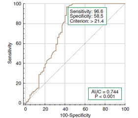
Актуальність. Незважаючи на прогрес у розумінні патогенезу та лікуванні діабетичної ретинопатії (ДР) та діабетичного макулярного набряку (ДМН) за цукрового діабету 2-го типу (ЦД2), визначення специфічних і чутливих біомаркерів є важливим для прогнозування та ранньої діагностики. Мета дослідження: встановити роль L-селектину у розвитку діабетичного макулярного набряку при діабетичній ретинопатії і цукровому діабеті 2-го типу. Матеріали та методи. Дослідження включало 124 пацієнти (124 ока) із ЦД 2-го типу, у яких за Міжнародною клінічною шкалою тяжкості ДР Американської академії офтальмології (2002 рік) виявлена легка (29 очей, 1-ша група), помірна або тяжка (35 очей, 2-га група) непроліферативна ДР та проліферативна ДР (31 око, 3-тя група); контрольну групу становили 29 пацієнтів без ЦД. Усім пацієнтам були виконані загальноприйняті офтальмологічні дослідження, а також спектральнодоменна оптична когерентна томографія (ОКТ) з визначенням наявності та класифікації ДМН за ОКТ-критеріями: збільшення товщини сітківки за нормативною базою даних, наявність інтраретинальної рідини, а також додаткові параметри: центральна товщина сітківки (ЦТС, мкм) та макулярний об’єм (МО). Уміст L-селектину у крові визначали імуноферментним методом (Invitrogen ThermoFisher Scientific, СШA). Для статистичних досліджень використано програмні пакети MedStat і MedCalc v.15.1 (MedCalc Software bvba). Результати. Уміст L-селектину у сироватці крові пацієнтів з ДР та ЦД2 був вірогідно збільшений у всіх групах (у 1-й групі у 2,0 раза, у 2-й — у 2,3 раза і у 3-й — у 3,2 раза порівняно з контролем; p < 0,05) та корелював з показниками, що відображали тяжкість порушення вуглеводного обміну (вмістом глюкози та глікованого гемоглобіну), та товщиною сітківки. При розподілі за наявністю ДМН уміст L-селектину був вищим тільки при помірній НПДР і не відрізнявся при інших стадіях ДР. Наявність патогенетичного зв’язку збільшення вмісту L-селектину з розвитком ДМН було підтверджено у регресійному аналізі: виявлено зростання ризику виникнення ДМН із зростанням вмісту L-селектину (ВШ = 1,09; 95% ВІ 1,05–1,14 на кожну одиницю зростання, нг/мл). Висновки. Результати дослідження підтвердили сучасну концепцію щодо значення L-селектину як одного з ключових біомаркерів запалення, що відіграють роль у розвитку ДР і ДМН при ЦД2.
Background. Despite progress in understanding the pathogenesis and treatment of diabetic retinopathy (DR) and diabetic macular edema (DME) in type 2 diabetes mellitus (T2DM), the identification of specific and sensitive biomarkers is important for prognosis and early diagnosis. The purpose of the study is to clarify the role of L-selectin in the development of diabetic macular edema in diabetic retinopathy and type 2 diabetes mellitus. Materials and methods. The study included 124 patients (124 eyes) with T2DM who had mild (29 eyes, group 1), moderate or severe (35 eyes, group 2) non-proliferative DR and proliferative DR (31 eyes, group 3) according to the International clinical scale of DR severity of the American Academy of Ophthalmology (2002); the control group consisted of 29 individuals without diabetes. All patients underwent standard ophthalmological examinations, as well as spectral domain optical coherence tomography (OCT) with the determination of the presence and classification of DME according to OCT criteria: an increase in retinal thickness according to the normative database, the presence of intraretinal fluid, and additional parameters: central retinal thickness (μm) and macular volume. The content of L-selectin in the blood was determined by the enzyme-linked immunosorbent assay (Invitrogen Thermo Fisher Scientific, USA). MedStat and MedCalc v.15.1 software packages (MedCalc Software bvba) were used for statistical research. Results. The content of L-selectin in the blood serum of patients with DR and T2DM was significantly increased in all groups (in group 1 by 2.0 times, in the second one by 2.3 times and in the third one by 3.2 times compared to the controls; p < 0.05) and correlated with indicators reflecting the severity of carbohydrate metabolism disorder (glucose and glycated hemoglobin content) and retinal thickness. When dividing by the presence of DME, the content of L-selectin was higher only in moderate non-proliferative DR and did not differ in other stages of DR. The presence of a pathogenetic relationship between an increase in the content of L-selectin and the development of DME was confirmed in regression analysis: an increase in the risk of DME with an increase in the content of L-selectin was found (odds ratio = 1.09; 95% confidence interval 1.05–1.14 for each unit of growth, ng/ml). Conclusions. The results of the study confirmed the modern concept of the importance of L-selectin as one of the key biomarkers of inflammation that play a role in the development of DR and DME in T2DM.
діабетичний макулярний набряк; діабетична ретинопатія; цукровий діабет 2-го типу; L-селектин, прогноз
diabetic macular edema; diabetic retinopathy; type 2 diabetes; L-selectin; prognosis
Для ознакомления с полным содержанием статьи необходимо оформить подписку на журнал.
- Teo Z.L., Tham Y.C., Yu M., Chee M.L., Rim T.H., Cheung N., et al. Global Prevalence of Diabetic Retinopathy and Projection of Burden through 2045: Systematic Review and Meta-ana–lysis. Ophthalmology. 2021 Nov. 128(11). 1580-1591. http://doi.org/10.1016/j.ophtha.2021.04.027.
- Wong T.Y., Cheung C.M., Larsen M., Sharma S., Simó R. Diabetic retinopathy. Nat. Rev. Dis. Primers. 2016 Mar 17. 2. 16012. http://doi.org/10.1038/nrdp.2016.12.
- Kirthi V., Nderitu P., Alam U., Evans J.R., Nevitt S., Malik R.A., et al. The prevalence of retinopathy in prediabetes: A systematic review. Surv. Ophthalmol. 2022 Sep-Oct. 67(5). 1332-1345. http://doi.org/10.1016/j.survophthal.2022.04.002.
- Hainsworth D.P., Bebu I., Aiello L.P., Sivitz W., Gubitosi-Klug R., Malone J., et al. Diabetes Control and Complications Trial (DCCT)/Epidemiology of Diabetes Interventions and Complications (EDIC) Research Group. Risk Factors for Retinopathy in Type 1 Dia–betes: The DCCT/EDIC Study. Diabetes Care. 2019 May. 42(5). 875-882. http://doi.org/10.2337/dc18-2308.
- Lu J., Ma X., Zhang L., Mo Y., Ying L., Lu W., et al. Glycemic variability assessed by continuous glucose monitoring and the risk of diabetic retinopathy in latent autoimmune diabetes of the adult and type 2 diabetes. J. Diabetes Investig. 2019 May. 10(3). 753-759. http://doi.org/10.1111/jdi.12957.
- Lin K.Y., Hsih W.H., Lin Y.B., Wen C.Y., Chang T.J. Update in the epidemiology, risk factors, screening, and treatment of diabetic retinopathy. J. Diabetes Investig. 2021 Aug. 12(8). 1322-1325. http://doi.org/10.1111/jdi.13480.
- Song J., Chen S., Liu X., Duan H., Kong J., Li Z. Relationship between C-Reactive Protein Level and Diabetic Reti–nopathy: A Systematic Review and Meta-Analysis. PLoS One. 2015 Dec 4. 10(12). e0144406. http://doi.org/10.1371/journal.pone.0144406.
- Tawfik A., Mohamed R., Elsherbiny N.M., DeAngelis M.M., Bartoli M., Al-Shabrawey M. Homocysteine: A Potential Biomarker for Diabetic Retinopathy. J. Clin. Med. 2019 Jan 19. 8(1). 121. http://doi.org/10.3390/jcm8010121.
- Xu J., Chen L.J., Yu J., Wang H.J., Zhang F., Liu Q., Wu J. Involvement of Advanced Glycation End Products in the Pathogenesis of Diabetic Retinopathy. Cell Physiol. Biochem. 2018. 48(2). 705-717. http://doi.org/10.1159/000491897.
- Mogilevskyy S.Yu., Panchenko Iu.O., Ziablitsev S.V. New risk factors for post-surgical recurrent diabetic maculopathy in type 2 diabetes mellitus. J. Оphthalmol. (Ukraine). 2019. 5. 9-17. http://doi.org/10.31288/oftalmolzh20195917.
- Nomura S., Omoto S., Yokoi T., Fujita S., Ozasa R., Eguchi N., Shouzu A. Effects of miglitol in platelet-derived microparticle, adiponectin, and selectin level in patients with type 2 diabetes mellitus. Int. J. Gen. Med. 2011. 4. 539-45. http://doi.org/10.2147/IJGM.S22115.
- Rahman I., Collado Sánchez A., Davies J., Rzeniewicz K., Abukscem S., Joachim J., et al. L-selectin regulates human neutrophil transendothelial migration. J. Cell Sci. 2021 Feb 8. 134(3). jcs250340. http://doi.org/10.1242/jcs.250340. Erratum in: J. Cell Sci. 2022 Oct 15. 135(20). PMID: 33408247; PMCID: PMC7888707.
- Mastej K., Adamiec R. Stezenie sL-selektyny w surowicy krwi oraz ekspresja L-selektyny na powierzchni leukocytów chorych z cukrzyca typu 2 [Serum level of sL-selectin and leukocyte surface expression of L-selectin in patients with type 2 diabetes]. Przegl Lek. 2011. 68(3). 140-5. Polish. PMID: 21812228.
- Newton V.L., Guck J.D., Cotter M.A., Cameron N.E., Gardiner N.J. Neutrophils Infiltrate the Spinal Cord Parenchyma of Rats with Experimental Diabetic Neuropathy. J. Diabetes Res. 2017. 2017. 4729284. http://doi.org/10.1155/2017/4729284.
- Pawłowski P., Urban M., Peczyńska J. Czy ekspresja L-selektyny moze być wczesnym markerem nadciśnienia tetniczego i mikroangiopatii w przebiegu cukrzycy typu 1 u młodocianych pacjentów? [Could the expression of L-selectin be an early marker of arterial hypertension and microangiopathy in the course of type 1 diabetes mellitus in juvenile patients?]. Endokrynol. Diabetol. Chor. Przemiany Materii Wieku Rozw. 2005. 11(3). 147-52. Polish. PMID: 16232368.
- Early Treatment Diabetic Retinopathy Study Research Group. Grading diabetic retinopathy from stereoscopic color fundus photographs — an extension of the modified Airlie house classification: ETDRS report № 10. Ophthalmology. 2020 Apr. 127(4S). 99-119. http://doi.org/10.1016/j.ophtha.2020.01.030.
- Гур’янов В.Г., Лях Ю.Є., Парій В.Д., Короткий О.В., Чалий О.В., Чалий К.О., Цехмістер Я.В. Посібник з біостатистики. Аналіз результатів медичних досліджень у пакеті EZR (R-statistics). Київ: Вістка, 2018. 208 с.
- Karadayi K., Top C., Gülecek O. The relationship between soluble L-selectin and the development of diabetic retinopathy. Ocul. Immunol. Inflamm. 2003 Jun. 11(2). 123-9. http://doi.org/10.1076/ocii.11.2.123.15920.
- MacKinnon J.R., Knott R.M., Forrester J.V. Altered L-selectin expression in lymphocytes and increased adhesion to endothelium in patients with diabetic retinopathy. Br. J. Ophthalmol. 2004 Sep. 88(9). 1137-41. http://doi.org/10.1136/bjo.2003.040329.
- Bartol-Puyal F.A., Isanta C., Verdes G., Ruiz-Moreno Ó., Calvo P., Pablo L. Influence of inflammatory plasma biomarkers on choroidal thickness in type 2 diabetes mellitus. Eur. J. Ophthalmol. 2023 Jan. 33(1). 468-482. http://doi.org/10.1177/11206721221124691.