
Журнал «Боль. Суставы. Позвоночник» Том 12, №4, 2022
Вернуться к номеру
Вплив альфакальцидолу на регенерацію кістки у щурів старечого віку
Авторы: Климовицький Ф.В. (1), Климовицький В.Г. (1), Дєдух Н.В. (2)
(1) — Донецький національний медичний університет, м. Лиман, Україна
(2) — ДУ «Інститут геронтології імені Д.Ф. Чеботарьова НАМН України», м. Київ, Україна
Рубрики: Ревматология, Травматология и ортопедия
Разделы: Клинические исследования
Версия для печати
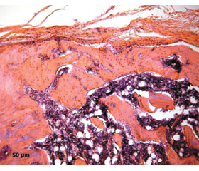
Актуальність. Серед препаратів, які мають плейотропну дію на кісткову тканину, особливу увагу привертає альфакальцидол — попередник активної форми D-гормона. Існують дослідження, у яких виявлено відмінні особливості впливу альфакальцидолу на регенерацію кістки за умов остеопорозу та в молодих тварин. Мета дослідження: вивчити особливості регенерації кістки після лікування альфакальцидолом у тварин старечого віку. Матеріали та методи. Щурам віком 24 місяці у ділянці метадіафіза моделювали транскортикальний дефект від латерального до медіального відділу стегнової кістки. Лікування альфакальцидолом проводили з 2-ї доби та протягом 10-ї та 30-ї діб до виведення тварин з експерименту. Проведено гістологічне дослідження регенерації кістки з морфометричним аналізом тканин у ділянках мозолі. Результати. На 10-ту добу після лікування альфакальцидолом тварин старечого віку в ділянці дефекту розташовувалася сполучна тканина, остеоїд та грубоволокниста трабекулярна кістка з високою щільністю остеобластів. На 30-ту добу у тварин, які отримували лікування, кісткова тканина регенерату мала пластинчасту будову й була зрілою порівняно з тваринами без лікування. У ділянці губчастої кістки була розташована сітка новоутворених кісткових трабекул. Деструктивні прояви навколо дефекту знижувалися у тварин, які отримували альфакальцидол. У тварин без лікування не виявлено щільного кісткового зрощення з фрагментами кортекса, трабекули губчастої кістки були переривчасті. У латеральній частині регенерату площа кісткової тканини була зменшена. Висновки. Лікування тварин альфакальцидолом прискорює формування кісткової тканини в дефекті та зменшує деструктивні прояви навколо дефекту порівняно з тваринами, яких не лікували.
Background. Among the drugs that have a pleiotropic effect on bone, attention is drawn to alfacalcidol – the precursor of the active form of D-hormone. There are studies that have revealed distinctive features of the effect of alfacalcidol on bone regeneration in osteoporosis and in young animals. The purpose was to study bone regeneration after treatment with alfacalcidol in senile animals. Materials and methods. A transcortical defect from the lateral to the medial femur was modeled in rats aged 24 months in the area of the metadiaphysis. A treatment with alfacalcidol was carried out from the 2-nd day and for 10 and 30 days before the animals was withdrawn from the experiment. Histological study of bone regeneration with morphometric analysis of tissues in the areas of callus had been performed. Results. On the 10-th day of the treatment with alfacalcidol in senile animals, connective tissue, osteoid and coarse-fibrous trabecular bone with a high density of osteoblasts were located in the defect area. On the 30-th day in treated animals, the bone tissue of the callus was mature; its area was larger than in untreated animals. In the area of cancellous bone there is a network of newly formed bone trabeculae. Destructive manifestations around the defect were reduced in animals treated with alfacalcidol. In untreated animals there was no dense bone fusion with cortex fragments; trabeculae of cancellous bone were intermittent. In the lateral part of the callus, the area of bone tissue was reduced compared to the medial part. Conclusions. Treatment of animals with alfacalcidol accelerates the formation of bone tissue in the defect and reduces destructive manifestations around the defect compared to untreated animals.
старечі щури; кістка; регенерація; гістологія; альфакальцидол
senile rats; bone; regeneration; histology; alfacalcidol
Для ознакомления с полным содержанием статьи необходимо оформить подписку на журнал.
- Meyer R.A., Tsahakis P.J., Martin D.F., et al. Age and ovariectomy impair both the normalization of mechanical properties and the accretion of mineral by the fracture callus in rats. J. Orthop. Res. 2001. 19(3). 428-435. doi: 10.1016/S0736-0266(00)90034-2.
- Borgiani E., Figge C., Kruck B. et al. Age-Related Changes in the Mechanical Regulation of Bone Healing Are Explained by Altered Cellular Mechanoresponse. Journal of Bone and Mineral Research (JBMR). 2019. 34(10). 1923-1937. doi: 10.1002/jbmr.3801.
- Pollock F.H., Maurer J.P., Sop A. et al. Humeral shaft fracture healing rates in older patients. Orthopedics. 2020. 43(3). 168-172. doi: 10.3928/01477447-20200213-03.
- Korzh N.A., Dedukh N.V., Horidova L.D. et al. Alfacalcidol in bone regeneration. Orthopedics, traumatology and prosthetics. 2013. 1. 73-83. doi: 10.15674/0030-59872013173-83.
- Liu D., Qin H., Yang J., et al. Different effects of Wnt/β-catenin activation and PTH activation in adult and aged male mice metaphyseal fracture healing. BMC Musculoskelet Disord. 2020 Feb 19. 21(1). 110. doi: 10.1186/s12891-020-3138-3.
- Gibon E., Lu L.Y., Nathan K., Goodman S.B. Inflammation, ageing, and bone regeneration. J. Orthop. Translat. 2017 Jul. 10. 28-35. doi: 10.1016/j.jot.2017.04.002.
- Gruber R., Koch H., Doll B.A., Tegtmeier F., Einhorn T.A., Hollinger J.O. Fracture healing in the elderly patient. Exp. Gerontol. 2006 Nov. 41(11). 1080-93. doi: 10.1016/j.exger.2006.09.008.
- Newman H., Shih Y.V., Varghese S. Resolution of inflammation in bone regeneration: From understandings to therapeutic applications. Biomaterials. 2021. 277. 121114. doi: 10.1016/j.biomaterials.2021.121114.
- Chen C.H., Wang L., Tulu U.S. et al. An osteopenic/osteoporotic phenotype delays alveolar bone repair. Bone. 2018. 112. 212-219. doi: 10.1016/j.bone.2018.04.019.
- Gurlek A., Pittelkow M.R., Kumar R. Modulation of growth factor/cytokine synthesis and signaling by 1alpha, 25-dihydroxyvitamin D(3): implications in cell growth and differentiation. Endocr. Rev. 2002 Dec. 23(6). 763-786. doi: 10.1210/er.2001-0044.
- European Convention for the protection of vertebrate animals used for experimental and other scientific purposes. Strasburg. 1986. ETS No. 123. Available from: https://rm.coe.int/168007a67b.
- Law of Ukraine On Protection of Animals from Cruelty. Information of the Verkhovna Rada of Ukraine 2006. № 27. Аrt. 230. Available from: https://zakon.rada.gov.ua/laws/show/3447-15#Text.
- Nair A.B., Jacob S. A simple practice guide for dose conversion between animals and human. J. Basic. Clin. Pharm. 2016 Mar. 7(2). 27-31. doi: 10.4103/0976-0105.177703.
- Sarkisov D.S., Perova Yu.L. Microscopic technique. М.: Medicine, 1996. 542.
- Avtandilov G.G. Medical morphometry: [manual]. M.: Medicine, 1990. 384.
- Ansari M. Bone tissue regeneration: biology, strategies and interface studies. Prog. Biomater. 2019 Dec. 8(4). 223-237. doi: 10.1007/s40204-019-00125-z.
- Aspenberg P. Special Review: Accelerating fracture repair in humans: a reading of old experiments and recent clinical trials. Bonekey Rep. 2013 Jan. 9. 2. 244. doi: 10.1038/bonekey.2012.244.
- Gao Y., Liu X., Gu Y. et al. The Effect of Bisphosphonates on Fracture Healing Time and Changes in Bone Mass Density: A Meta-Analysis. Front. Endocrinol. 2021. 12. 688269. doi: 10.3389/fendo.2021.688269.
- Klontzas M.E., Kenanidis E.I., Mac Farlane R.J. et al. Investigational drugs for fracture healing: preclinical & clinical data. Expert Opin. Investig. Drugs. 2016. 25(5). 585-96. doi: 10.1517/13543784.2016.1161757.
- Поворознюк В.В., Климовицкий Ф.В., Дедух Н.В. Морфологические особенности регенерации транскортикального метадиафизарного дефекта при лечении животных альфакальцидолом. Український медичний альманах. 2013. 16(1). 35-39. Режим доступу: http://nbuv.gov.ua/UJRN/Uma_2013_16_1_10.
- van Driel M., Pols H.A., van Leeuwen J.P. Osteoblast differentiation and control by vitamin D and vitamin D metabolites. Curr. Pharm. Des. 2004. 10(21). 2535-2555. doi: 10.2174/1381612043383818.