Інформація призначена тільки для фахівців сфери охорони здоров'я, осіб,
які мають вищу або середню спеціальну медичну освіту.

Підтвердіть, що Ви є фахівцем у сфері охорони здоров'я.


UkrainePediatricGlobal

UkrainePediatricGlobal

Журнал «Здоровье ребенка» Том 16, №2, 2021

Вернуться к номеру

Ендоскопічно-морфологічна характеристика верхніх відділів шлунково-кишкового тракту в дітей із харчовою гіперчутливістю

Авторы: L.M. Bubyr, T.O. Kryuchko, I.M. Nesina, B.M. Filenko, T.O. Pedchenko
Ukrainian Medical Stomatological Academy, Poltava, Ukraine

Рубрики: Педиатрия/Неонатология

Разделы: Клинические исследования

Версия для печати


Резюме

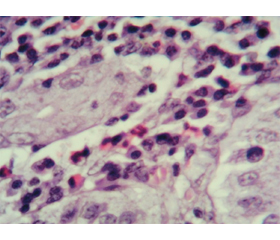
Актуальність. Останнім часом в педіатричній практиці все більше уваги приділяється вивченню алергічних уражень різних відділів шлунково-кишкового тракту, а ряд невирішених питань стосовно проблем своєчасної діагностики та лікування еозинофільних уражень органів травлення обумовлює актуальність цієї тематики. Мета: вивчити ендоскопічно-морфологічні зміни верхніх відділів шлунково-кишкового тракту в дітей із гастроінтестинальними симптомами харчової гіперчутливості. Матеріали та методи. У дослідженні взяли участь 34 дитини віком від 6 до 15 років із клінічними ознаками ураження верхніх відділів травного тракту на тлі реакцій харчової гіперчутливості. Залежно від рівня загального IgE учасники дослідження були розподілені на дві групи. Першу групу становили 18 дітей з IgE-незалежними алергічними реакціями на їжу (Me (Q1-Q3) 35,0 (28,0–77,5)). До другої групи було включено 16 пацієнтів з IgE-обумовленими алергічними проявами харчової гіперчутливості (Me (Q1-Q3) 240,5 (158,0–475,8)). Для досягнення поставленої мети всім дітям було проведено фіброезофагогастродуоденоскопію з подальшим забором матеріалу та наступною морфологічною оцінкою біоптатів. Результати. Згідно з результатами ендоскопічного обстеження пацієнтів, структура уражень верхніх відділів шлунково-кишкового тракту залежно від переважання IgE-незалежних чи IgE-опосередкованих гастроінтестинальних симптомів харчової гіперчутливості не мала статистично значущих відмінностей. Морфологічні характеристики мали певні відмінності в досліджуваних групах й характеризувалися превалюванням еозинофільної інфільтрації в дітей з IgE-обумовленими реакціями харчової гіперчутливості. Висновки. В ендоскопічній картині дітей із гастроінтестинальними проявами харчової алергії переважають ізольовані ураження шлунка. Морфологічні ж ознаки хронічного гастриту характеризуються змінами покривного епітелію за рахунок вираженої поліморфноклітинної запальної інфільтрації у власній пластинці слизової оболонки з переважанням лімфоцитів і плазмоцитів та порівняно меншою кількістю нейтрофілів, еозинофілів та макрофагів. При цьому активність еозинофільного запального процесу була більш вираженою в групі дітей з IgЕ-обумовленими реакціями харчової гіперчутливості (r = 0,652; р < 0,01).

Background. In pediatric practice, increasing attention has been recently paid to the study of allergic di­sorders in various parts of the gastrointestinal tract, and a number of unresolved issues regarding the timely diagnosis and treatment of eosinophilic lesions of the digestive system determines the relevance of this topic. The purpose of the research was to study the endoscopic and morphological changes of the upper gastrointestinal tract in children with gastrointestinal symptoms of food hypersensitivity. Materials and methods. The study enrolled 34 children aged from 6 to 15 years with clinical signs of disorders of the upper digestive tract against the background of food hypersensitivity reactions. Depen­ding on the level of total IgE, study participants were divided into two groups. The first group consisted of 18 children with IgE-independent allergic reactions to food (Me (Q1-Q3) 35.0 (28.0–77.5)). The second group included 16 patients with IgE-induced allergic manifestations of food hypersensitivity (Me (Q1-Q3) 240.5 (158.0–475.8)). To achieve the aim of the research, all children underwent fibroesophagogastroduodenoscopy with subsequent sampling and morphological evaluation of biopsy specimen. Results. According to the results of endoscopic examination of patients, the structure of lesions of the upper gastrointestinal tract depending on the predominance of IgE-independent or IgE-mediated gastrointestinal symptoms of food hypersensitivity did not have statistically significant differences. Morphological characteristics had some differences in the study groups and were represented by the prevalence of eosinophilic infiltration in children with IgE-induced food hypersensitivity reactions. Conclusions. The isolated gastric lesions prevailed in the endoscopic presentation of children with gastrointestinal manifestations of food allergy, and morphological signs of chronic gastritis are characterized by changes in the surface epithelium due to pronounced polymorphonuclear inflammatory infiltration in the lamina propria with a predominance of lymphocytes, neutrophils and plasma cells. The degree of activity of the eosinophilic inflammatory process was more pronounced in the group of children with IgE-induced food hypersensitivity reactions (r = 0.652; p < 0.01).


Ключевые слова

харчова алергія; патологія шлунково-кишкового тракту; еозинофільний гастроентерит; діагностика; морфологічні ознаки

food allergy; pathology of the gastrointestinal tract; eosinophilic gastroenteritis; diagnosis; morphological signs

Background

In pediatric practice, increasing attention has been recently paid to the study of allergic disorders in various parts of the gastrointestinal tract [1–3]. According to an estimate from the national administrative database of the United States of America, published in the Journal of Pediatric Gastroenterology and Nutrition in 2016, the prevalence of eosinophilic gastritis amounted to 6.3 cases, and eosinophilic gastroenteritis — 8.4 cases per 100 thousand population. Among children of early and preschool age, eosinophilic gastroenteritis had the highest prevalence, whereas the incidence of eosinophilic gastritis was higher in older children [4]. Diagnosis of eosinophilic gastroenteritis is a rather time-consuming task for practical medicine due to the lack of uniform standardized diagnostic algorithms and specific clinical criteria, as well as insignificant information value of allergological examination due to IgE-independent mechanism of inflammation, which, in turn, requires the search for additional immunological studies to verify the diagnosis of food allergies [5].
The pathophysiological mechanisms underlying different types of food allergy reactions have significant differences [6, 7]. Eosinophilic gastroenteritis is an idiopathic inflammatory lesion of the gastrointestinal tract that belongs to a mixed type of allergic reactions due to IgE- and cell-mediated (IgE-independent) mechanisms and is characterized by eosinophilic infiltration of the stomach and/or intestine. At the present stage, IgE-mediated allergic reactions induced by dietary proteins remain the least studied. One of the approaches to their understanding is the stimulation of T helper (Th) mechanisms with the participation of effector immune cells — lymphocytes, which cause the production of proinflammatory cytokines — interleukins (IL) 4, 5, 13 and promote the migration of eosinophils into the stomach and intestine. The IgE-independent immune response may also be accompanied by mast cell activation [8].
As can be seen from Fig. 1, during the initial entry of a food allergen into the body, for example, orally, by inhalation, or percutaneously, there is a presentation of antigenic determinants of the allergen by sensitive cells (e.g., dendritic). This, in turn, activates T helpers and stimulates the functional activity of B lymphocytes in local lymph nodes, as well as the triggering mechanisms of immune tolerance or sensitization. Dendritic cells play a leading role in this process. During tolerance formation, an important place belongs to retinoic acid and the production of anti-inflammatory cytokines IL-10 and transforming growth factor β, which affects the formation of tolerogenic Treg cells. In turn, B cells can produce sIgA, which neutralizes antigenic epitopes. In the absence of conditions necessary for the induction of tolerance, sensitization by food allergens occurs. This releases epithelial cytokines: IL-33, thymic stromal lymphopoietin, and IL-25, which indirectly stimulate the differentiation of Th0 by Th2 type. The formed Th2 cells secrete a number of cytokines of allergic inflammation, such as IL-5, which is responsible for eosinophil сhemotaxis, as well as IL-4 and IL-13. Specific IgEs bind to high-affinity Fcɛ receptor I (FcƐRI) on the surfaces of basophils and mast cells. Upon further contact with food allergen, activation and degranulation of these cells occur with the release of anti-inflammatory factors. Studies show that a food allergen entering the circulatory system leads to the formation of an immune response. This ensures the implementation of IgE-induced allergic immune response [6, 9, 10].
Histopathological examination of gastric and duodenal samples plays a crucial role in the diagnosis of eosinophilic gastroenteritis and should be based on the detection of eosinophilic mucosal infiltration. There is no generally accepted threshold for the number of eosinophils because under normal conditions they are present in the wall of the gastrointestinal tract. The most acceptable indicator is the number of eosinophils exceeding 20 cells in the field of view in at least one biopsy sample. However, these data do not take into account the age of the patient and the segment of the digestive tract, whereof the biopsy was taken.
Thus, given the complex cascade of immune responses in patients with a suspected food allergy, morphological evaluation of gastrointestinal mucosa biopsy specimen remains the gold standard, along with clinical history and provocation and elimination measures, which determines the relevance of the study of endoscopic and morphological characteristics, manifestations of allergic hypersensitivity against the background of food consumption.
The purpose of our research was to study the endoscopic and morphological changes of the upper gastrointestinal tract in children with gastrointestinal symptoms of food hypersensitivity.

Materials and methods

The study enrolled 34 children aged from 6 to 15 years with clinical signs of disorders of the upper gastrointestinal tract, represented by gastroesophageal reflux disease, chronic gastritis, and duodenitis against the background of food hypersensitivity reactions, with the voluntary consent obtained from parents. The basis for the selection of respondents in this study was the presence of significant clinical and anamnestic data that suggest the suspicion of food allergies and, therefore, the positive results of allergy testing. Depending on the level of total IgE, participants were divided into two groups. The first group consisted of 18 children with IgE-independent allergic reactions to food (Me (Q1-Q3) 35.0 (28.0–77.5)). The second group included 16 patients with IgE-induced allergic manifestations of food hypersensitivity (Me (Q1-Q3) 240.5 (158.0–475.8)). To achieve the aim of the research, all children underwent fibroesophagogastroduodenoscopy with subsequent sampling from different parts of the esophagus, stomach, and duodenum, as well as morphological evaluation of biopsy specimen. Fixation of the obtained material was performed in a 10% solution of neutral formalin, followed by histological sample preparations according to standard methods. Sections were stained with hematoxylin and eosin. The number of eosinophils was calculated based on the threshold diagnostic criteria depending on the digestive tract segment, specified in the works of many researchers [11–14] in five random high-power fields (HPF), which is a morphological criterion of an allergic reaction. Microscopic examination of stained histological specimens was performed on a light microscope MICROmed XS-3320 (Ningero Sheng Heng Optics and Electronics Co.) with 100×, 200×, 400× magnification.
Statistical analysis of the results was made using Microsoft Excel 2010 software. The arithmetic mean of the number of eosinophils and their standard deviation were calculated. The relationship between eosinophil count and serum IgE levels was assessed using nonparametric Spearman’s and Kendall’s correlation coefficients (r). The relationship between the studied parameters was identified as weak, moderate, and strong if the correlation coefficient was in the range of 0.01–0.29, 0.3–0.69, and 0.70–1.0, respectively. All indicators were considered reliable at a value of p ≤ 0.05.

Results and discussion

The results of endoscopic examination of patients in our study (Table 1) show that the structure of lesions of the upper gastrointestinal tract depending on the predominance of IgE-independent or IgE-mediated gastrointestinal symptoms of food hypersensitivity had no statistically significant differences in the study groups. The distribution of endoscopic forms such as gastritis and duodenitis was almost uniform and was 94.4 % in group 1 versus 100 % in group 2 in the first case, and 44.4 against 50.0 % with inflammatory changes in the duodenum. It should be noted that among patients with IgE-independent manifestations of food hypersensitivity, in some cases there were erosive (5.5 %) and ulcerative (5.5 %) defects of the gastric and duodenal mucosa, which is comparable to data from other researches [15]. The predominance of destructive changes in the mucous membrane can be explained by the long-term course of gastrointestinal symptoms, which causes the chronicity of the inflammatory process through the links of cell-mediated mechanisms of allergy formation [16]. In addition, patients of this population had more frequent, as compared to children of group 2, motor dysfunctions of the cardiac and pyloric regions of the stomach, manifested by gastroesophageal (16.7 %) and duodenogastric (44.4 %) reflux. A characteristic feature of children with IgE-induced food hypersensitivity reactions was a higher level of Helicobacter pylori infection — 75 against 61.1 % in patients from group 1, which does not contradict the data from other researches [17]. Analysis of the literature provides data on the prevalence of organic changes in the gastrointestinal tract among 46–78 % of children with food allergies, whereas functional disorders are diagnosed in almost all patients [18, 19].
It should be noted that endoscopic examination did not reveal any inflammatory changes of the esophageal mucosa in the form of esophagitis. However, in 3 (16.7 %) children of group 1 and 1 patient (6.3 %) with elevated levels of total IgE, we registered a decrease in esophageal pH < 4, which in combination with typical clinical symptoms gave grounds for the diagnosis of gastroesophageal reflux disease without esophagitis. According to the results of microscopic studies, eosinophilic infiltration in the mucous membrane of the esophagus was not detected. This can be explained by the structural and morphological features of the mucous membrane of this organ and the impossibility of sampling, taking both the mucous and submucosal layers for the entire thickness.
Microscopic examination of gastric biopsy specimen in group 1 of children showed preservation of the structure of the mucous membrane with dystrophic changes of the epithelium and signs of chronic inflammation. The lamina propria was represented by loose connective tissue, which sporadically had moderate cellular infiltration, represented by lymphocytes, neutrophils, macrophages, plasma cells, and eosinophils. Some eosinophils were in a state of degranulation (Fig. 2).
Besides, it should be noted that in 17 (94.4 %) cases, a ratio of neutrophilic granulocytes, plasma cells, and lymphocytes was uniform, which probably indicates the activation of the inflammatory process. This was clinically manifested by signs of exacerbation of chronic gastritis. Calculation of the median distribution of eosinophils, which were a part of the inflammatory infiltrate of the gastric mucosa in children with non-IgE-mediated food hypersensitivity reactions, showed their wide fluctuations in the antrum with an average value of 20.94 ± 1.82 in HPF.
Although the obtained data were significantly lower compared to the children with elevated IgE levels, the study found a moderate correlation (Fig. 3) between total IgE content and the number of eosinophils in the inflammatory infiltrate of the gastric mucosa (r = 0.522; p < 0.01), which can be explained by the fact that these changes belong to the delayed-type allergic reactions and by the absence of lower normative values of immunoglobulin E in children of different ages.
Microscopic examination of the duodenal mucosa in this group of patients showed the preservation of its histological structure with a clear differentiation of the mucosa and submucosal layer. The mucous membrane had a well-defined villous surface. Intestinal villi were represented by finger-shaped protrusions of loose connective tissue, covered with a single layer of prismatic epithelium. A thin layer of mucus was determined on the surface of the mucous membrane. It should be noted that only 8 (44.4 %) children of this group demonstrated severe signs of an active inflammatory process, manifested by severe edema of the intestinal villi with varying degrees of inflammatory cell infiltration, represented by lymphocytes, plasma cells, and segmental leukocytes with a small number of eosinophils. In addition, the same changes were observed in the submucosal base of these patients, but they were more pronounced compared to the mucous membrane, and the cellular composition included a higher number of eosinophils (Fig. 4).
The number of eosinophils in biopsy samples of the duodenal mucosa in this group of patients averaged 22.56 ± 3.04 in HPF (min = 4.0; max = 54.0). In addition, there is an uneven distribution in the mucous membrane with their predominant accumulation in some areas and the absence or single location in others. The obtained data indicate no statistically significant correlation between total IgE levels and eosinophil counts in the duodenal mucosa of patients with IgE-independent gastrointestinal symptoms of food hypersensitivity (r = 0.291; p > 0.05).
Microscopic examination of gastric biopsy samples in the group of children with IgE-induced manifestations of food hypersensitivity showed preservation of the histological structure of the stomach, but all (100 %) subjects had the signs of chronic inflammation. Comparing the histological presentation in the previous group, we have found more pronounced signs of pathological changes. There was no clear differentiation of cell pools as a result of the absence of mucous secretion in the apical part and the presence of dystrophic changes. On the surface of the mucous membrane, the mucus layer was either thin or completely absent. Desquamation of single epithelial cells or their layers was observed.
In all cases, there was a pronounced polymorphonuclear inflammatory infiltration in the lamina propria with a predominance of lymphocytes and plasma cells and a relatively small number of neutrophils, eosinophils, and macrophages. Diffuse inflammatory infiltration spread to the entire thickness of the lamina propria (Fig. 5). In addition, there was an uneven repletion of blood vessels, predominantly venous-capillary plethora with perivascular edema and diapedetic hemorrhages.
It should be noted that in some cases there was a pronounced eosinophilic infiltration with focal accumulation and severe edema of the stroma in these areas. Determination of the number of eosinophils in the five high-power fields showed that in patients with IgE-mediated allergic reactions to food it averaged 39.06 ± 3.91 in HPF, with significant fluctuations in some patients, which was statistically significantly higher than in group 1 (p < 0.01). There was a moderate correlation (Fig. 6) between total IgE and the number of eosinophils in the gastric mucosa (r = 0.652; p < 0.01).
When studying duodenal biopsy specimen in this group of patients, we found that only half of the children had severe signs of inflammation. Histologically, this was manifested by deformation of the microvilli of the mucous membrane due to severe swelling of their stroma. They were thickened and had folds and protrusions, but their epithelial lining was preserved. A large amount of mucus containing desquamated enterocytes was detected on the surface of the villi and in the depth of the crypts. The stroma of microvilli had a pronounced inflammatory infiltration with predominance of lymphocytes and plasma cells and an admixture of a relatively small number of other cells. Dystrophic processes were observed in the acinar parts of the duodenal glands. Among the inflammatory infiltrate cells, eosinophils constituted a significant number. In addition, eosinophilic granules were detected extracellularly (Fig. 7).
The number of eosinophils in duodenal biopsy specimen of patients with IgE-induced gastrointestinal symptoms was 1.5 times higher compared to samples in the group of children with food hypersensitivity with normal IgE levels (p < 0.05) and averaged 33.63 ± 4.28 in HPF (min = 11.0; max = 61.0). However, no correlation was demonstrated between total IgE and the number of duodenal eosinophils in patients of this group, which can be explained by the lower proportion of verified duodenitis in both groups.
Thus, the results of the endoscopic examination demonstrated that the lesion of the gastric and duodenal mucosa had no specific signs that would indicate food hypersensitivity. These data are confirmed by studies of some authors and indicate the need for histological examination [20]. However, despite this fact, there is a constant search for macroscopic changes in the mucous membrane of the gastrointestinal tract and their justification in terms of clinical, laboratory, and microscopic data, and it is proposed to take into account a number of features that are statistically significant [21, 22]. Therefore, currently, mucosal biopsy is a reliable diagnostic method for the detection of characteristic changes and eosinophilic infiltration [23]. At the same time, this method is traumatic and has disadvantages for its application in pediatric practice. Therefore, the search for and optimization of the diagnosis of food allergies in children according to clinical and laboratory data is an urgent problem of today, which requires a comprehensive study [24]. We attempted to determine the relationship between total IgE levels and the severity of eosinophilic infiltration in the gastric and duodenal mucosa. The identified changes indicate that in children with IgE-mediated allergic reactions to food, along with pronounced morphological signs of chronic gastritis, there was a greater number of eosinophils, which were a part of the inflammatory infiltrate in the gastric antrum and the duodenum, compared to patients without IgE-mediated allergic reactions. There is a relationship of varying strength between the severity of eosinophilic infiltration in the gastric and duodenal mucosa and the level of total IgE.

Conclusions

The isolated gastric lesions prevailed in the endoscopic presentation of children with gastrointestinal manifestations of food allergy, whereas the morphological signs of chronic gastritis are characterized by changes in the surface epithelium due to pronounced polymorphonuclear inflammatory infiltration in the lamina propria with a predominance of lymphocytes, neutrophils and plasma cells. The degree of activity of the eosinophilic inflammatory process was more pronounced in the group of children with IgE-induced food hypersensitivity reactions (r = 0.652; p < 0.01). At the same time, for the final verification of allergic gastrointestinal lesions in patients with normal IgE levels, a combined histological evaluation of biopsy samples with immunological markers of atopy is required taking into account clinical and anamnestic data.
Conflicts of interests. Authors declare the absence of any conflicts of interests and their own financial interest that might be construed to influence the results or interpretation of their manuscript.
Funding. This article has been written within the research work of the Department of Pediatrics 2 of the Ukrainian Medical Stomatological Academy “Study of pathogenetic mechanisms of the most common childhood illnesses, optimization of diagnosis and treatment” 2017–2021 (state registration number 0117U004683).
 
Received 27.01.2021
Revised 10.02.2021
Accepted 20.02.2021

Список литературы

  1. Уманец Т.Р., Шадрин О.Г., Клименко В.А. и др. Основные положения руководства по ведению больных с аллергией к коровьему молоку. Часть І. Эпидемиология и аллергены коровьего молока. Современная педиатрия. 2015. 1(65). 16-22.
  2. Охотникова Е.Н., Черныш Ю.Р. Гастроинтестинальная аллергия в практике педиатра и детского аллерголога. Здоровье ребeнка. 2015. 4. 15-21.
  3. Боброва В.І., Богданова Т.А. Гастроінтестинальна гіперреактивність та її значення у формуванні хронічного алергічного гастриту у дітей. Современная педиатрия. 2016. 1(73). 127-129. doi: 10.15574/SP.2016.73.127.
  4. Jensen E.T., Martin C.F., Kappelman M.D., Dellon E.S. Prevalence of eosinophilic gastritis, gastroenteritis, and colitis: estimates from a national administrative database. J. Pediatr. Gastroenterol. Nutr. 2016. 62(1). 36-42. doi: 10.1097/MPG.0000000000000865.
  5. Захарова И.Н., Бережная И.В. Пищевая аллергия у детей: с чем связан ее рост? Медицинский совет. 2018. 17. 156-162.
  6. Muraro A., Werfel T., Hoffmann-Sommergruber K., Roberts G., Beyer K., Bindslev-Jensen C. et al. EAACI food allergy and anaphylaxis guidelines: diagnosis and management of food allergy. Allergy. 2014 Aug. 69(8). 1008-25. doi: 10.1111/all.12429.
  7. Makarova S.G., Namazova-Baranova L.S., Vishneva E.А., Ereshko O.А., Gordeeva I.G. Gastrointestinal food allergy in children. Voprosy sovremennoi pediatrii — Current Pediatrics. 2017. 16(3). 202-212. doi: 10.15690/vsp.v16i3.1730.
  8. Зайцева Ю.Г., Халева Е.Г., Жданова М.В., Новик Г.А. Не-IgE-зависимая пищевая аллергия у детей. Лечащий врач. 2018. 4. 31-34.
  9. Betty van Esch. Cows milk allergy. Avoidance versus tolerance: new concepts for allergy management. Utrecht, the Netherlands. 2011. 1. 10.
  10. Sharon Chinthrajah R., Hernandes Joseph D., Boyd Scott D., Galli Stephen J., Nadeau Kari C. Molecular and cellular mechanisms of food allergy and food tolerance. J. Allergy Clin. Immunol. 2016 Apr. 137(4). 984-997.
  11. Lwin T., Melton S., Genta R. Eosinophilic gastritis: histopathological characterization and quantification of the normal gastric eosinophil content. Mod. Pathol. 2011. 24. 556-63.
  12. Sunkara T., Rawla P., Yarlagadda K.S., Gaduputi V. Eosinophilic gastroenteritis: diagnosis and clinical perspectives. Clin. Exp. Gastroenterol. 2019. 12. 239-53.
  13. Reed C., Woosley J.T., Dellon E.S. Clinical characteristics, treatment outcomes, and resource utilization in children and adults with eosinophilic gastroenteritis. Dig. Liver Dis. 2015. 47(3). 197-201.
  14. Черниш Ю.Р., Охотнікова О.М. Клінічні прояви гастроінтестинальної форми харчової алергії у дітей і підходи до її діагностики. Здоров’я дитини. 2017. 12(5). 611-622.
  15. Богданова Т.А., Березенко В.С., Пьянкова А.В., Гурьева О.В., Голованенко Г.Н. Особливості перебігу хронічного гастродуоденіту на тлі харчової гіперчутливості у підлітків. Современная педиатрия. 2018. 3(91). 54-58. doi: 10.15574/SP.2018.91.54.
  16. Шадрін О.Г., Задорожна Т.Д., Гайдучик Г.А., Арчакова Т.М., Місник В.П. Клініко-морфологічні особливості алергічного ентероколіту в дітей раннього віку. Патологія. 2019. 16(2). 238-244.
  17. Сорокман Т.В., Попелюк О.-М.В., Лозюк І.Я. Особливості перебігу поєднаної патології верхніх відділів шлунково-кишкового тракту та алергодерматозів у дітей. Здоров’я дитини. 2017. 12(3). 324-328.
  18. Боткина А.С. Пищевая аллергия у детей: современный взгляд на проблему. Лечащий врач. 2012. 6. 24-26.
  19. Абатуров А.Е., Кайдашев И.П., Никулина А.А. и др. Противовоспалительные эффекты пробиотической терапии хронических гастродуоденитов у детей. Здоровье ребенка. 2020. 15(5). 287-293.
  20. Pesek R.D., Reed C.C., Collins M.H., Muir A.B., Fulkerson P.C., Menard-Katcher C. et al. Association between endoscopic and histologic findings in a multicenter retrospective cohort of patients with non-esophageal eosinophilic gastrointestinal disorders. Dig. Dis. Sci. 2020. 65. 2024-2035.
  21. Fujiwara Y., Tanoue K., Higashimori A., Nishida Y., Maruyama M., Itani S. et al. Endoscopic findings of gastric lesions in patients with eosinophilic gastrointestinal disorders. Endosc. Int. Open. 2020. 8(12). 1817-1825.
  22. Ashitani K., Tsuzuki Y., Yamaoka M., Ohgo H., Ichimura T., Kusano T. et al. Endoscopic features and diagnostic procedures of eosinophilic gastroenteritis. Internal Medicine. 2019. 58(15). 2167-2171.
  23. Calvani M., Anania C., Cuomo B., D’Auria E., Decimo F., Indirli G.C. et al. Non-IgE- or mixed IgE/non-IgE-mediated gastrointestinal food allergies in the first years of life: old and new tools for diagnosis. Nutrients. 2021. 13(1). 226. doi: 10.3390/nu13010226.
  24. Kryuchko T.O., Bubyr L.M., Nesina I.M., Tkachenko O.Y., Izmailova O.V., Poda O.A. et al. Ways of optimizing the diagnostics of food allergies in children based on the clinical and immunological criteria. Wiad. Lek. 2020. 73(10). 2255-2260.

Вернуться к номеру