Інформація призначена тільки для фахівців сфери охорони здоров'я, осіб,
які мають вищу або середню спеціальну медичну освіту.

Підтвердіть, що Ви є фахівцем у сфері охорони здоров'я.


UkraineNeuroGlobal


UkraineNeuroGlobal

Международный неврологический журнал Том 16, №1, 2020

Вернуться к номеру

Концентрація алюмінію та свинцю в сироватці крові потерпілих на мирній території з черепно-мозковою травмою легкого й середнього ступеня тяжкості в гострому і проміжному періодах

Авторы: M.G. Semchyshyn, V.M. Shevaga
Danylo Halytsky Lviv National Medical University, Lviv, Ukraine

Рубрики: Неврология

Разделы: Клинические исследования

Версия для печати


Резюме

Актуальність. Патогенез черепно-мозкової травми (ЧМТ) багатогранний і значною мірою зумовлений розвитком ендогенної інтоксикації, що веде до накопичення в організмі різних проміжних і кінцевих продуктів метаболізму, у тому числі й токсичних металів. Розуміння основних шляхів патогенезу інтоксикації алюмінієм і свинцем дозволить розробити методи ранньої діагностики, а також дієві заходи з профілактики та лікування. Мета роботи: вивчити зміни показників алюмінію й свинцю в сироватці крові потерпілих мирної території з ЧМТ легкого й середнього ступеня тяжкості в гострому й проміжному періодах. Матеріали та методи. Проведене визначення вмісту алюмінію й свинцю в сироватці крові в 283 потерпілих мирної території з ЧМТ методом атомно-абсорбційної спектрофотометрії з використанням повітряно-ацетиленового полум’я. Результати. З огляду на наявні зміни вмісту токсичних мікроелементів у сироватці крові потерпілих мирної території з ЧМТ легкого й середнього ступеня тяжкості в гострому й проміжному періодах можна стверджувати про наявність у них метаболічних порушень у тканині мозку, пригнічення ендотеліальної функції судин і посилення процесів апоптозу. На нашу думку, у вказаних пацієнтів ендотеліальна дисфункція може розглядатися як маркер судинної патології. Висновки. Таким чином, дослідження вмісту токсичних мікроелементів у сироватці крові потерпілих мирної території з ЧМТ легкого й середнього ступеня тяжкості в гострому й проміжному періодах можуть бути використані як діагностичний і прогностичний критерії перебігу травматичної хвороби головного мозку та в експертних цілях, а вивчення основних шляхів патогенезу алюмінієвої та свинцевої інтоксикації дозволить розробити методи їх ранньої діагностики, дієві заходи з профілактики й лікування.

Актуальность. Патогенез черепно-мозговой травмы (ЧМТ) многогранный и в значительной степени обусловлен развитием эндогенной интоксикации, которая приводит к накоплению в организме различных промежуточных и конечных продуктов метаболизма, в том числе и токсических металлов. Понимание основных путей патогенеза интоксикации алюминием и свинцом позволит разработать методы ранней диагностики, а также действенные меры по профилактике и лечению. Цель работы: изучить изменения показателей алюминия и свинца в сыворотке крови потерпевших на мирной территории с ЧМТ легкой и средней степени тяжести в остром и промежуточном периодах. Материалы и методы. Проведено определение содержания алюминия и свинца в сыворотке крови у 283 потерпевших на мирной территории с ЧМТ методом атомно-абсорбционной спектрофотометрии с использованием воздушно-ацетиленового пламени. Результаты. С учетом очевидных изменений содержания токсических микроэлементов в сыворотке крови потерпевших на мирной территории с ЧМТ легкой и средней степени тяжести в остром и промежуточном периодах можно говорить о наличии у них метаболических нарушений в ткани мозга, угнетении эндотелиальной функции сосудов и усилении процессов апоптоза. По нашему мнению, у указанных пациентов эндотелиальная дисфункция может рассматриваться как маркер сосудистой патологии. Выводы. Таким образом, исследования содержания токсических микроэлементов в сыворотке крови потерпевших на мирной территории с ЧМТ легкой и средней степени тяжести в остром и промежуточном периодах могут быть использованы в качестве диагностического и прогностического критериев течения травматической болезни головного мозга и с экспертными целями, а изучение основных путей патогенеза алюминиевой и свинцовой интоксикации позволит разработать методы их ранней диагностики, действенные меры по профилактике и лечению.

Background. Pathogenesis of the traumatic brain injury is multifaceted and is largely dependent on the development of endogenous intoxication, which leads to the accumulation of various intermediate and end products of metabolism, including toxic metals, in the body. Understanding the basic pathways of the pathogenesis of aluminium and lead intoxication will allow developing the early diagnostic methods as well as effective preventive and treatment measures. The paper aimed to study changes in blood serum concentration of aluminium and lead in persons from the peaceful territory with mild to moderate traumatic brain injury in acute and intermediate periods. Materials and methods. The blood serum concentration of aluminium and lead in 283 people of the peaceful territory with traumatic brain injury of mild to moderate severity in acute and intermediate periods was determined by atomic absorption spectrophotometry using an air-acetylene flame. Results. Given the changes in the content of toxic trace elements in the blood serum of people of the peaceful territory with traumatic brain injury of mild to moderate severity in the acute and intermediate periods, it can be argued that they have metabolic disorders in the brain tissue, inhibited endothelial function of the vessels and intensification of the apoptosis processes. In our opinion, endothelial dysfunction in these people of the peaceful territory may be considered as a marker of vascular pathology. Conclusions. Thus, the study of toxic trace elements in the blood serum of people of the peaceful territory with traumatic brain injury of mild to moderate severity in the acute and intermediate periods can be used as diagnostic and prognostic criteria for the course of traumatic brain disease and for expert purposes, and the study of the major pathways of pathogenesis of aluminium and lead intoxication will allow developing the methods for their early diagnosis, effective measures of prevention and treatment.


Ключевые слова

алюміній; свинець; черепно-мозкова травма

алюминий; свинец; черепно-мозговая травма

aluminium; lead; brain injury

Introduction

The problem of brain injury is one of the significant multidisciplinary problems today. During the last years, we observe the increase of neurotraumatism which is caused by urbanization, the development of transport, the accele-rated rhythm of life. The mild to moderate traumatic brain injury holds the first place in the structure of craniocerebral traumas. Disability caused by the brain injury of the mild to moderate severity is more often resulted from cognitive and mental disorders, rather than neuropathological ones, which in their turn determinate the problem of social adaptation and the working rehabilitation of a person, who suffered from the brain injury [7, 8, 11, 14].
The toxic metals, as well as technological pollution, play a significant role in the pathogenesis of the brain injury, influence the vessels and tissue of the brain especially. As a result of absorption and accumulation in the body, they block the activity of cellular enzymes and their systems, provoke serious alterations in cellular metabolism and cause intoxication [6]. The clinical manifestations of intoxication and its spectrum depend on the concentration of metal and the duration of exposure. Even a slight degree of aluminium and lead intoxication is severe and may trigger mutation processes [1, 4]. Mild intoxication is often latent and its manifestation may be unspecific. Aluminium and lead get accumulated in the vessel wall and develop vasotoxic effect [9, 13]. This state increases the generation of the reactive oxygen species and develops oxidant stress that elevates the accumulation of free iron in the body. It negatively impacts the exchange of nitrogen oxide that leads to modifying the functional quality of the vessel wall (endothelial dysfunction) [2, 10]. Understanding the primary mechanisms of pathogenesis of aluminium and lead intoxication will enable the elaboration of the methods for early diagnosis, prophylaxis and treatment [3, 12].
Purpose of the study: to study the changes in blood serum concentration of aluminium and lead in people of the peaceful territory with mild to moderate traumatic brain injury in acute and intermediate periods.

Materials and methods

We have investigated three groups of people of the peaceful territory with brain injury: concussion — 143 patients, mild brain contusion — 119 patients, moderate brain contusion — 21 patients. The comparison group included 20 healthy people aged 18–50 years. The investigated group covered 191 men and 92 women aged 20–55 years. The blood for analysis was taken from the vein on an empty stomach on the days 1–2, 3–5, 7–10, 14–21, in 1 and 3 months after the trauma. The blood serum concentration of aluminium and lead was determined by the method of atomic absorption spectrophotometry along with using an air-acetylene flame [5]. Statistical processing helped to calculate an arithmetical average, average quadratic deviation, an average error, significant difference concerning the results of the investigation and the Student’s t-test.

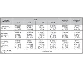
Results and discussion

The Tables 1, 2 demonstrate the data of the blood serum concentration of aluminium and lead in people of the peaceful territory with brain injury and healthy people.
According to the data in Table 1, the blood serum concentration of aluminium in patients with concussion and brain contusion of mild severity did not differ from this of the control group (p > 0.05) in acute and intermediate periods. However, in patients with moderate brain contusion, the level of aluminium probably, not significantly (p > 0.05) was higher than in the comparison group on days 1–2, 3–5, 7–10, 14–21 and a month after the trauma. Three months later, the content of aluminium normalized and reached the level of the control one (p > 0.05).
In the group of patients with concussion, the highest level of aluminium was determined on day 3–5 and was doubtful compared to the content on day 1–2 (p > 0.05). On day 7–10, the concentration of aluminium decreased and the change was doubtful compared to this one on day 1–2 (p > 0.05), however, it was significantly lower on day 3–5 (p < 0.05). The content of aluminium was significantly lower on days 1–2, 3–5, 7–10 (p < 0.05) and on day 14–21. One month after the trauma, the level of aluminium slightly increased and remained significantly lower than it was on days 1–2, 3–5, 7–10 (p < 0.05) and significantly higher compared to the day 14–21 (p < 0.05). Three months later, the content of aluminium was significantly lower compared to the days 1–2, 3–5, 7–10 and one month after the trauma (p < 0.05), but remained doubtful compared with the level on day 14–21 (p > 0.05).
In patients with the mild brain contusion, the highest level of aluminium was on day 3–5 and significantly higher than on day 1–2 (p < 0.05). On day 7–10, the content of the metal decreased in the blood serum of patients and was doubtful compared to the level on day 1–2 (p > 0.05), and significantly lower than on day 3–5 (p < 0.05). On day 14–21, the concentration of aluminium kept on reducing and was significantly lower than on days 1–2, 3–5, 7–10 (p < 0.05). One month later, the content of aluminium did not change compared to the level on days 14–21 and was doubtfully changeable (p > 0.05), but significantly lower than on days 1–2, 3–5, 7–10 (p < 0.05). Three months later, the concentration of metal decreased more and was significantly lower than the content on days 1–2, 3–5, 7–10, 14–21 and a month after the trauma (p < 0.05).
In the group of patients with the brain contusion of moderate severity, the highest level of aluminium was determined on days 14–21. On days 3–5, the level of aluminium increased and remained significantly higher than on days 1–2 (p < 0.05). On days 7–10, its concentration further increased in the blood serum of patients and was significantly higher than on days 1–2, and doubtfully higher than on days 3–5 (p > 0.05). On days 14–21, the level of aluminium was significantly higher than on days 1–2, 3–5, 7–10 (p < 0.05). One month later, the content of metal reduced and remained doubtfully higher than on days 1–2 (p > 0.05) and significantly lower than on days 3–5, 7–10, 14–21 (p < 0.05). Three months later, the content of aluminium was significantly lower than on days 1–2, 3–5, 7–10, 14–21 and a month later (p < 0.05).
We have revealed a significant difference between the content of aluminium in the blood of patients with the concussion and the brain contusion of mild severity on days 1–2, 3–5, 7–10, after 1 and 3 months (p1 < 0.05), however, the data were doubtful on days 14–21 (p1 > 0.05). In the patients with the concussion and the brain contusion of moderate severity, the significant difference was concerning to the level on days 1–2, 3–5, 7–10, 14–21 and a month after the trauma (p2 < 0.05), while after 3 months, the difference was doubtful (p2 > 0.05). In the groups of patients with the brain contusion of mild and moderate severity, the significant changes were determined during all the periods of observation on days 1–2, 3–5, 7–10, 14–21, after 1 and 3 months (p3 < 0.05).
All the three investigated groups of patients with the brain injury of mild to moderate severity in acute and intermediate periods showed no significant difference in the level of lead in comparison with the control group (p > 0.05) (Table 2).
We have identified essential discrepancy in the concentration of lead between patients with the concussion and those with the brain contusion of mild severity on days 1–2, 3–5, 7–10, 14–21 (p1 < 0.05); and after 1 and 3 months, the data were doubtful (p1 > 0.05). In patients with concussion in comparison with those with the brain contusion of moderate severity, the blood serum content of metal was reliable on days 1–2, 3–5, 7–10, 14–21 and after 1 month (p2 < 0.05), after 3 months the data were doubtful (p2 > 0.05). In the groups with the brain contusion of mild to moderate severity, the lead levels were doubtful within all the periods of observation (p3 > 0.05).

Conclusions

1. The increase of aluminium in patients with brain injury may lead to the changes of rheological properties of blood, provoke the development of early atherosclerosis and inhibit the activity of enzymes in the energetic exchange of cells.
2. The change of the content of toxic microelements tracing in the blood serum after a brain injury causes metabolic problems in the brain tissue, the synthesis of pro-inflammatory enzymes, the inhibition of endothelial functions in the vessels and the intensification of apoptosis.
3. The endothelial dysfunction is considered a marker of the vascular pathology, essential for progressing and clinical manifestations in the patients with the brain injury of mild to moderate severity in acute and intermediate periods.
4. The study of the main ways of pathogenesis of aluminium and lead intoxication can enable to elaborate the methods for early diagnosis, prophylaxis and treatment.

Prospects for further investigation

The identified changes in the content of toxic microele-ments in the blood serum in patients with brain injury of mild to moderate severity in acute and intermediate periods will specify and explain some aspects of pathogenesis and clinical manifestations in this disease. This may have an impact on diagnosis and prediction, it is also important for determining the necessity of a differential therapeutic approach.
Conflicts of interests. Authors declare the absence of any conflicts of interests and their own financial interest that might be construed to influence the results or interpretation of their manuscript.

Список литературы

1. Бойченко П.К., Лузин В.И., Хорькова Е.А. и др. Влияние алюминия на обмен биоминералов. Укр. морфологический альманах. 2006. 4(2). 120-121.

2. Волошин П.В., Малахов В.А., Завгородняя А.Н. Эндотелиальная дисфункция при церебральной патологии. Харьков: Украина, 2006. 92 с.

3. Кудрин А.В., Громова О.А. Микроэлементы в неврологии М.: ГЭОТАР-Медиа, 2006. 304 с.

4. Зербино Д.Д., Соломенчук Т.М., Поспишиль Ю.А. Свинец — этиологический фактор поражения сосудов: основные доказательства. Архив патологии. 1997. 59(1). 9-12.

5. Текуцкая Е.Е., Софьина Л.И., Бендер Л.В., Онищенко Н.П. Методы и практика контроля содержания тяжелых металлов в биосредах. Гигиена и санитария. 1999. 4. 72-74.

6. Штабский Б.М., Гжегоцкий М.Р. Профилактическая токсикология и прикладная физиология: общность проблем и пути решения. Львов: НАУТИЛУС, 2003. 342 с.

7. Тайцлин В.И. Закрытая черепно-мозговая травма и ее последствия. Междунар. мед. журнал. 2002. 1–2. 58-62.

8. Педаченко Є.Г., Шлапак І.П., Гук А.П., Пилипенко М.М. Черепно-мозкова травма: сучасні принципи невідкладної допомоги: навч.-метод. посібник. К.: ВАРТА, 2007. 312 с.

9. Апихтіна О.Л., Коцюруба А.В., Коркач Ю.П. та ін. Вазотоксична дія свинцю: роль порушень в системі оксиду азоту. Укр. журн. з проблем медицини праці. 2007. 3(11). 56-62.

10. Зенков Н.К., Ланкин В.З., Меншикова Е.Б. Окислительный стресс. Биохимический и патофизиологический аспекты. М.: МАИК «Наука-Интерпериодика», 2001. 343 с.

11. Живолупов С.А., Самарцев И.Н., Коломенцев С.В. Опыт применения препарата «Аксамон» в комплексной терапии больных с черепно-мозговыми травмами в восстановительном периоде. Российский национальный конгресс «Человек и лекарство». М., 2009. 100-101.

12. Davidson T., Ke Q., Costa M. Transport of Toxic Metals by Molecular/Ionic Mimicry of Essential Compounds. In Handbook on the toxicology of metals. 3rd ed. Ed. by Nordberg G.F. et al. Acad. Press, London New York Tokyo, 2007. P. 79-84.

13. Prozialeck W.C., Edwards J.R., Nebert D.W. et al. The Vascular System as a Target of Metal Toxicity. Toxicological Sciences. 2008. 102(2). 207-18.

14. Rapoport M.J., McCullagh S., Shammi P. et al. Cognitive impairment associated with major depression following mild and moderate traumatic brain injury. The Journal of Neuropsychiatry and Clinical Neurosciences. 2005. 17. 61-5.


Вернуться к номеру