Статтю опубліковано на с. 20-24
Вступ
Серед усіх різновидів переломів верхньої кінцівки найбільш поширеними є переломи променевої кістки в типовому місці (ППКТМ). У процентному співвідношенні вони становлять 40–45 % від усіх переломів скелета [6].
У травматології на сучасному етапі спостерігається тенденція охоплення оперативним методом хворих із ППКТМ при їх лікуванні [1]. Це значною мірою
зумовлено створенням високотехнологічних фіксаторів [2, 8]. Проте застосування модерних засобів для консервативної та оперативної фіксації фрагментів при ППКТМ не завжди забезпечує добрий результат лікування [3]. Для з’ясування причин такого стану методики лікування ППКТМ повинні всебічно вивчатися за допомогою ефективних методологічних підходів.
Мета дослідження: проаналізувати сучасні тенденції консервативних і оперативних методів лікування ППКТМ та визначити фактори, що впливають на їх результати.
Матеріали та методи
Ми провели системний аналіз (СА) основних факторів консервативного й оперативного методів лікування ППКТМ. Саме СА, імплікативне мислення при плануванні методики лікування, оперативного втручання, способу фіксації відламків дозволять розібратися в гносеології проблеми, дають можливості з’ясувати вплив засобів фіксації на зрощення кісткових фрагментів, пов’язати наслідок фіксації з багатьма чинниками, що її визначають [4].
Проведений аналіз доступних літературних джерел, методик консервативного лікування, застосування LCP-пластин, апаратів зовнішньої фіксації при ППКТМ. Виявлені причини можливих ускладнень і фактори, що впливають на результати лікування цих пошкоджень. Вивчені також наявні рентгенограми та медична документація, історії хвороби 55 пацієнтів, у яких виявлені ускладнення і негативні результати застосування сучасних засобів для остеосинтезу. Аналізувались біомеханічна обґрунтованість застосування обраної методики лікування, конструкції фіксатора для конкретного перелому, її вплив на зрощення відламків. При цьому важливе значення має правильна оцінка характеру перелому. Іноді вона можлива тільки при вивченні даних комп’ютерної, магнітно-резонансної томографії. Зокрема, при переломі Бартона [5] у 2 випадках для уточнення характеру перелому була проведена комп’ютерна томографія. Вивчалась динаміка розвитку мозолі. Відмічалась залежність її величини від якості репозиції відламків, стабільності фіксації, що визначалась особливостями конструкції фіксатора. Для консервативного методу лікування важливе значення має стан гіпсової пов’язки, своєчасність рентгенологічного контролю. Значна увага приділялась взаємодії окремих елементів, простежувались динаміка процесу, вплив нових важливих чинників на зв’язок структури і функції, коректність методологічного підходу, раціональність тактичних рішень.
При аналізі консервативних методик вивчалися можливість закритої репозиції фрагментів, в яких біомеханічних умовах вона проходить, а також засоби для стабілізації відламків. Зокрема, розглядалися: гіпсова пов’язка, пластиковий гіпс (скотчкаст), ортези та інші синтетичні пов’язки. Вивчалася взаємодія пов’язки з фрагментом — вона в основному визначає ті процеси, що відбуваються на лінії контакту відламків, і забезпечує кінцевий результат лікування перелому.
Завдяки СА, що розглядає елементи та підсистеми у взаємозв’язку, орієнтовані на досягнення кінцевої мети, можна розробити профілактичні заходи та прогнозувати наслідок загоювання ППКТМ [2, 4]. На основі СА можлива розробка нових, нестандартних підходів у лікуванні ППКТМ, що складатиме нове знан-
ня та нову парадигму цієї проблеми.
Аналізу піддавалися також багато інших об’єктивних і суб’єктивних факторів, що мають вплив на кінцевий результат. Зокрема, фіксувалася відповідність лікування конкретного випадку рекомендованій методиці, а також техніка виконання оперативного втручання, правильність проведення післяопераційного періоду, що є визначальними в отриманні доброго кінцевого результату.
Результати та обговорення
Перелом променевої кістки більше характерний для жінок і людей похилого віку. Сама по собі променева кістка відносно тонка, а з віком цей показник зменшується в рази. Часті випадки перелому такого типу можна пояснити особливостями її анатомічної структури: дистальний кінець променевої кістки має найменшу товщину кортикального шару. Цей перелом зустрічається у двох видах: згинальний перелом (Сміта) і розгинальний (Коллеса). Причиною екстензійного пошкодження є падіння пацієнта на витягнуту руку при тильній флексії кисті. У дорослих людей при цьому може спостерігатися вколочений перелом без явного зміщення. Флексійний перелом виникає при падінні на руку в положенні долонної флексії кисті. При цьому площина зламу відбувається спереду назад і знизу вгору. Дистальний фрагмент зміщується проксимально в долонну сторону. Таке пошкодження зустрічається досить рідко, проте часто роблять помилку при іммобілізації, здійснюючи фіксацію як при розгинальному переломі.
При переломах зі зміщенням в ділянці променево-зап’ясткового суглоба визначається багнетоподібна деформація. На тильній стороні іноді вдається пропальпувати дистальний уламок, а на долонній — проксимальний. При переломах Сміта, навпаки, на тильній стороні — центральний, на долонній — периферичний. За відсутності зсуву деформація визначається тільки за рахунок гематоми. Пальпація особливо болюча на тильній стороні променевозап’ясткового суглоба на рівні перелому. Осьове навантаження викликає посилення болю в місці перелому. Перевіряти рухомість між відламками та кісткову крепітацію не варто.
Крім анамнезу та клінічної картини вирішальне значення для діагнозу ППКТМ мають рентгенограми у двох проекціях (прямій і бічній). Правильна оцінка характеру перелому, зміщення дистального уламку визначає подальшу тактику лікування. Слід пам’ятати, що суглобова фасетка головки ліктьової кістки розташована на 0,5–1 см проксимальніше суглобової поверхні променевої кістки, яка нахилена в долонну сторону під кутом 10°. Допускається горизонтальне її положення. Нормальний кут між суглобовою поверхнею променевої кістки і перпендикуляром до осі діафіза на прямій рентгенограмі (радіоульнарний кут) становить 30°. Зсув головки ліктьової кістки в дистальному напрямку і зміна радіоульнарного кута за відсутності репозиції зумовлюють обмеження ліктьового відведення кисті і ротаційних рухів передпліччя.
Неправильна інтерпретація рентгенограм визначає помилкову подальшу тактику, сприяє збільшенню кількості ускладнень і незадовільних результатів. Ми спостерігали 7 пацієнтів, у яких неправильна оцінка рентгенограм стала причиною нераціональної тактики лікування, що вплинула на кінцевий результат.
Здебільшого при ППКТМ репозиція фрагментів проводиться під місцевою анестезією. Для цього в місце перелому вводять 20 мл 1% розчину новокаїну або стільки ж 0,5% лідокаїну. У 3 пацієнтів при несвіжих випадках ми здійснювали репозицію під загальним знеболюванням. Вправити фрагменти можна при повноцінному знеболюванні та біомеханічно правильно проведеній репозиції. Для цього відламки необхідно спочатку роз’єднати шляхом посилення деформації та розтягнення. Більшість травматологів не зважають на цей важливий момент. Просте розтягнення з’єднаних фрагментів часто виявляється неефективним, особливо в несвіжих випадках.
Наприкінці репозиції при переломах Коллеса кисть фіксують у положенні достатнього долонного згинання й ульнарного відхилення. Після репозиції перелому Сміта гіпсова пов’язка стабілізує кисть у положенні тильної флексії. Для профілактики вторинного зміщення пов’язка повинна щільно облягати пошкоджений сегмент, але не здавлювати його. Це зменшує також частоту розвитку нейродистрофічного синдрому.
При осколковому ППКТМ після репозиції відламків у хворого Д. проведена фіксація їх спицями (рис. 1). Загалом така методика застосована нами в 4 випадках. Вона забезпечує мінімальну травматизацію пошкодженого сегмента [9].
Традиційно для виявлення вторинного зміщення на 8–10-й день проводяться контрольні рентгенограми. Проте вторинне зміщення може настати й пізніше. Ми спостерігали двох пацієнтів, у яких зміщення настало через три тижні після репозиції.
/22.jpg)
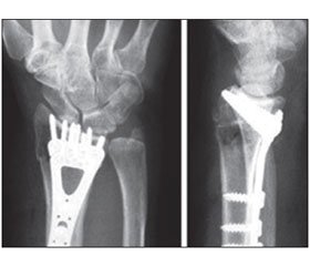
Практика показала, що останнім часом у таких випадках проводиться відкрита репозиція МОС LCP-пластинами [8]. Правильно сплановане оперативне втручання, анатомічна репозиція відламків забезпечили добрий результат у хворого П. (рис. 2). Проте при осколкових ППКТМ МОС LCP-пластиною провести досить складно, особливо у випадках несвіжих переломів. Дрібні відламки тяжко піддаються репозиції та фіксації пластиною з гвинтами. Переважно це закінчується розвитком артрозу. Таке ускладнення ми спостерігали у хворого М. (рис. 3). Найбільш оптимальним у цьому випадку був би остеосинтез апаратом зовнішньої фіксації [7]. Останній із позитивним результатом ми використали у 24 випадках. Саме позавогнищевий остеосинтез дозволив нам добитися доброго результату при переломі Бартона у хворого К. (рис. 4).
/22_2.jpg)
Перелом Бартона — це внутрішньосуглобовий перелом дистальної частини променевої кістки з вивихом у променевозап’ястковому суглобі [5]. При цьому частина суглобової фасетки в момент перелому розвертається на 180°, тому його ще називають «реверсний перелом», що відрізняє його від інших ППКТМ. Механізм травми — падіння на розігнутий та пронований променево-зап’ястковий суглоб із сильним ударом у дорзальну частину суглобової щілини. Закрита репозиція та утримання досягнутих результатів можливі тільки в поодиноких випадках. Останнім часом популярним стає відкрите вправлення з подальшою фіксацією LCP-пластинкою. Проте на практиці зафіксувати дрібний відламок практично неможливо. Ми вважаємо, що методом вибору в таких випадках є позавогнищевий остеосинтез із відкритою репозицією відламків і фіксацією їх спицями.
Саме така методика нами була застосована у хворого К. Після невдалої закритої репозиції накладено апарат Ілізарова (рис. 5). Апаратом здійснено розтягнення променевозап’ясткового суглоба, Z-подібним доступом по долонній поверхні правого променево-зап’ясткового суглоба, вздовж променевого згинача зап’ястка до 8,0 см оголено місце перелому. Виявлено, що частина суглобової фасетки розвернута на 180°. Проведені репозиція відламка фасетки та фіксація його 2 спицями Кіршнера (рис. 6).
У даній ситуації нами обрана найбільш оптимальна методика лікування перелому Бартона при дрібних відламках суглобової поверхні променевої кістки із реверсом. Ми вважаємо, що застосування LCP-пластини у таких випадках не показане.
Висновки
Таким чином, СА основних факторів консервативного й оперативного методів лікування ППКТМ дає можливість з’ясувати вплив засобів фіксації на зрощення кісткових фрагментів. Отримати добрий кінцевий результат можна, урахувавши багато об’єктивних і суб’єктивних чинників: ґрунтовний аналіз характеру лінії перелому, вибір найбільш оптимальної, біомеханічно обґрунтованої методики лікування та методично правильне її виконання.
Конфлікт інтересів. Автори заявляють про відсутність конфлікту інтересів при підготовці даної статті.
Список литературы
1. Андрейчин В.А. Системний аналіз оперативного методу лікування діафізарних переломів і фактори впливу на репаративну регенерацію / В.А. Андрейчин, П.І. Білінський // Травма. — 2014. — № 6. — С. 59-64.
2. Білінський П.І. Теорія і практика малоконтактного багатоплощинного стеосинтезу / П.І. Білінський. — К.: Макрос, 2008. — 375 с.
3. Ролік О.В. Післятравматичний нейродистрофічний синдром при переломах дистального метаепіфізу кісток передпліччя / О.В. Ролік, Т.С. Ганич, Г.І. Колісник // Ортопедия, травматология и протезирование. — 2004. — № 1. — С. 127-132.
4. Сименач Б.И. Фрактурология — некоторые аспекты теоретизации учения о переломах костей. Часть 2. Управление процессами репарации / Б.И. Сименач // Ортопедия, травматология и протезирование. — 2000. — № 4. — С. 105-117.
5. Baron J.A. Radial Differences in fracture risk / J.A. Baron, J.A. Barrett, D. Malenka // Epidemiology. — 2004. — № 5. — P. 42-47.
6. Gartland J.J. Evaluation of healed Colles’fractures / J.J. Gartland, C.W. Werley // J. Bone Jt. Surg. — 2002. — Vol. 33-A. — P. 895-907.
7. Ludvigsen T.S. External fixation versus percutaneus pinning for unstable colles fracture. Equal autcome in a randomized study of 60 patients / T.S. Ludvigsen, S. Johansen, S. Svenningsen // Acta Orthop. Scand. — 2007. — № 68. — P. 255-258.
8. McQueen M.M. The value of remapulating Colles fractures / M.M. McQueen, A. MacLaren, J. Chalmers // Jt. Bone Jt. Surg. — 2006. — Vol. 68-B, № 2. — P. 232-233.
9. Scheck M. Long-term follow up of treatment of comminuted fractures of distal end of the radius by transfixation with Kirschner wires and cast / M. Scheck // J. Bone Jt. Surg. — 2012. — Vol. 44-A. — P. 337-351.
/22.jpg)


/22_2.jpg)
/23.jpg)