
Журнал «Здоровье ребенка» (62) 2015. Тематический выпуск "Детская гастроэнтерология"
Вернуться к номеру
Clinical case of alagille syndrome
Авторы: Omelchenko E.V.(1), Єrmolaєv M.N.(2), Senatorova A.S.(1), Shipko A.F.(1), Omelchenko- Selyukova A.V. (1), Єrmolaєva M.M. (2), Levchenko Y.A.(2)
(1) — Kharkiv National Medical University
(2) — Kharkiv Regional Children’s Clinical Hospital, Kharkiv, Ukraine
Рубрики: Педиатрия/Неонатология
Разделы: Клинические исследования
Версия для печати
Alagille syndrome, also known as arteriohepatic dysplasia, is an autosomal dominant disorder that traditionally has been defined by a paucity of intrahepatic bile ducts, in association with 5 main clinical abnormalities: cholestasis, cardiac disease, skeletal abnormalities, ocular abnormalities, and a characteristic facial phenotype. Cholestasis is a direct consequence of the paucity of bile ducts. In addition to neonatal jaundice, features of this syndrome include the following: in the eye, posterior embryotoxon and retinal pigmentary changes; in the heart, pulmonic valvular stenosis as well as peripheral arterial stenosis; in the bones, abnormal vertebrae ('butterfly' vertebrae) and decrease in interpediculate distance in the lumbar spine; in the nervous system, absent deep tendon reflexes and poor school performance; in the facies, broad forehead, pointed mandible and bulbous tip of the nose and in the fingers, varying degrees of foreshortening. Alagille syndrome is transmitted in an autosomal dominant pattern of inheritance with incomplete penetrance. The syndrome was first described in 1969 by French paediatrician Daniel Alagille and his associates. Approximately two in every 100,000 children are born with Alagille syndrome. Most cases of the syndrome are caused by a mutation in a gene known as JAG1 , found on chromosome 20 (20p12.2). JAG1 controls the formation of (codes for) a protein called JAG1.
There is an own observation case. The patient B., 11 months was enrolled in the Kharkiv Regional Children’s Clinical Hospital. Parents have complained that a child had of intense yellow skin or mucous membranes with a greenish tinge, lethargy, vomiting, rectal prolapse.
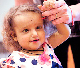
From the case history is known that at the age of 1, 5 months against the backdrop of persistent jaundice skin appear soft stools, ecchymosis. Cryptogenic hepatitis was diagnosed in hematology department. Child was transferred to the Regional Children's Infectious Hospital, where he remained for a month, were it was diagnosed as a congenital cytomegalovirus infection, hepatitis. Atresia of the intrahepatic bile ducts was excluded. Lethargy, drowsiness increased after 3 months, yellowness of the skin acquired a greenish tinge, appeared polyfecalia and clarification of feces. Conditions were very severe on admission to the gastroenterological department: malnutrition grade 2 (weight 7kg), general weakness, muscle hypotonia, delayed psycho-motor development (not sitting on their own). Patient had yellowness of the skin, mucous membranes and sclera with a greenish tint, pastose face, feet and legs. Phenotypic patient variables were triangular shaped face with a broad prominent forehead, deep-set eyes, flattened, "bird" nose and pointed chin, crooked V hands fingers. Heart sounds was rhythmic, loud systolic hum on an apex. Belly was increased in size, ascites. The liver is palpated for 3 cm by the midclavicular line, solid consistency, spleen up to 2.5 cm. There were colored stools, light yellow, polyfecalia, prolapse of the rectum and dark urine. Examination revealed cholestasis syndrome (cytolysis, hyperbilirubinemia, increasing of alkaline phosphatase). Differential diagnosis was held between congenital tyrosinemia, Baylera disease, incomplete biliary atresia, biliary cirrhosis secondary on the background of the transferred generalized CMV — infection, glycogen storage disease, peroxisome diseases and other aminoacidopathy, Konovalov-Wilson's disease, cystic fibrosis, deficiency of alpha-1-antitrypsin. Results of computer tomography of the lumbosacral spine were — CT signs of anomalies of the lumbosacral junction (lumbarization of L6, spina bifida L6), agenesis of the coccyx. Doppler echocardiography: left-right shunt diameter 4.8-5.5 mm in the central part of the interatrial septum. Conclusion of the liver biopsies was: Chronic hepatitis with smallnodullar cirrhosis. Liver biopsy with violation of tissue structure due to the formation of the set of false lobules, with giant metamorphosis hepatocytes. Lobules were separated by wide connective tissue septa with irregular mononuclear leukocyte infiltration. The combination of fetal malnutrition, hypoplasia of the intrahepatic bile ducts, congenital heart disease(atrial septal defect), biochemical markers of cholestasis, and changes in the musculoskeletal system (facial dysmorphia, signs of abnormal development of the lumbosacral junction (lumbarization of L6, spina bifida L6), agenesis of the coccyx, the backlog in physical development ware the basis of the diagnosis: Alagille sindrom.
Report of the transplant surgeon was — end-stage liver disease, intrahepatic cholestasis syndrome "Alagille." He recommended a liver transplantation. The deterioration of the child condition on the background of disease progression occurred in 2 months. The child became sharply adynamic, drowsiness, occasionally opened his eyes, joined disseminated intravascular coagulation syndrome, increased manifestations of cerebral, cardiovascular, respiratory failure. On the third day of hospitalization registered biological death.
Diagnostic of Alagille syndrome is based on the identification of the characteristic features of the phenotype and 2 or more typical anomalies and / or malformations of other organs and / or systems. Systemic lesions in Alagille syndrome complicate the course of the disease and make treatment difficult. The choice of surgical approach depends of the combined multiple malformations.
Список литературы
1. Degtyareva A.V. «Differentsial'naya diagnostika i printsipy etiopatogeneticheskogo lecheniya zabolevaniy pecheni i zhelchnykh putey u novorozhdennykh i detey rannego vozrasta» avtoreferat na soiskaniye nauchnoy stepeni doktora meditsinskikh nauk. Moskva 2010god .40s.
2. Fabris L., Cadamuro M., Gvido M., Spirli S, Fiorotto R., Colledan M., Torre G., Al'berti D., Sonzogni A., Okolicsanyi L., Strazzabosco M. Analiz pecheni remontu mekhanizmov v Alazhilya sindrom i biliarnoy atrezii pokazyvayet rol' dlya Notch signalizatsii. Am J Pathol. 2007; 171 (2): 641-653.
3. Hirschfield G. Novyye vzglyady na patogenez pervichnogo biliarnogo tsirroza pecheni i pervichnyy skleroziruyushchiy kholangit // Ils poslediplomnogo kursa «Kholestaticheskiye zabolevaniya pecheni i zhelchnykh protokov». — Berlin, 2011. — S. 3-69.
4. Kamath B., Mun'os P., Bab N., Beyker A. i dr. Prodol'noye issledovaniye, chtoby vyyavit' laboratornyye prediktory pecheni iskhoda zabolevaniya pri sindrome Alazhilya. J Pediatr Gastroenterol Nutr. 2010; 50 (5): 526-530.
5. Ling SC vrozhdennyye kholestaticheskogo sindromov: Chto proiskhodit, kogda deti vyrastayut? Mozhet J Gastroenterol. 2007; 21 (11): 743-751.
6. Raukh R. Hofbeck M., Zweier C., Koch A., Tsinka S., Trautmann, U., Khoyyer J., Kaulitts R., Zinger H., Raukh. Kompleksnyy analiz genotip-fenotip 230 patsiyentov s Fallo. J. Med. Zhene. 47: 321-331, 2010.