
Журнал «Здоровье ребенка» 2 (61) 2015
Вернуться к номеру
The onset and evolution characters of juvenile rheumatoid arthritis symptomatology during in the first year of its development.
Авторы: Lebets I.S.(1), Panko N.O.(2), Matvienko A.V.(1), Zaytseva E. N.(1), Shevchenko N.S.(1)
(1) — SI "Institute for Children and Adolescents Health Care of the NAMS of Ukraine"
(2) — V.Karazin Kharkiv National University
Рубрики: Педиатрия/Неонатология
Разделы: Клинические исследования
Версия для печати
Ключевые слова
children, early juvenile rheumatoid arthritis, symptoms, diagnosis.
Abstract. The article presents the data on early symptoms of juvenile rheumatoid arthritis (JRA) in comparison with the reactive arthritis (RA) signs. Description of the articular syndrome, analysis of the frequency of isolated joints affection and certain changes on the X-ray patterns are also given in the article. The designed algorithm is intended for differential diagnosis of juvenile rheumatoid and reactive arthrites.
Background. JRA remains so far a disease with unfavorable prognosis considering its course and consequences. From the conventional point of view this pathology is characterized by a steady development, invalidity of patients and their short life-span. Early diagnostic criteria for JRA have not been developed up to now, and existing criteria for RA in adults do not meet the age-related requirements for children. All the above defined the aim of our research: to estimate manifestations of JRA in children at its onset and during certain months during the first year of the disease development, as well as to identify the most informative early diagnostic signs of the pathology.
Materials and methods.
89 persons aged 2-18 years with clinical and sonographic signs of arthritis were examined in the in-patient clinic of the SI "Institute for Children and Adolescents Health Care of the NAMS”. Articular form of JRA was diagnosed in 38 patients, and RA diagnosis was verified in 51 patients (comparison group). Duration of both diseases by the time of initial examination did not exceed 12 months. The International Classification of Diseases, 10th revision, and special records for diagnosing cardiorheumatologic diseases in children (by order of the Ministry of Health, No. 362 of 2005) were used in our study. Clinical symptomatology was studied in our patients together with evaluation of functional capacity of the joints and a thorough examination of the locomotor system, the number of active joints according to the Ritchie index, as well as definition of pain intensity on the visual analogue scale (VAS). C-reactive protein (C-RP) level and rheumatoid factor were determined as well. Statistical analysis of the data obtained was carried out using the application package.
Results. Evaluation of clinical symptoms of the diseases under investigation has shown that poor health, increased fatigability, and general muscular asthenia are determined mainly in JRA (p <0.01), and their frequency and severity correspond to the pathological process activity. According to the degree of its activity, patients with JRA were distributed as follows: 66.3% of patients had a minimal degree, 24.7% - a moderate (second) degree, and 9% - a third degree. It has been registered that in RA its third degree is not recorded at all, and a second degree occurs not often (p <0.01). Attention should be focused on such an important for JRA diagnostic sign for as morning stiffness. It was determined in 78.9% of patients since the onset of the disease, in 31.6% of patients it remained till midday, in 21.1% it stayed for 1-2 hours, and in the rest morning stiffness duration was about 20 minutes. Certain torpidity concerning reverse manifestations of morning stiffness was registered during treatment of patients with JRA. In patients with RA morning stiffness signs were rare and short-lived, as compared with JRA.
A typical manifestation of both diseases was pain in the joints, prevailing in the morning hours. However, in JRA arthralgia persisted in most patients much longer and stayed even overnight. Higher rates of pain intensity have been revealed according to Haskinson’s VAS in patients with JRA, as compared with it in patients, suffering from RA (p<0,01).
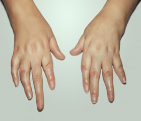
Clinical evaluation of the articular syndrome during the first year of the disease development has revealed polyarthritis prevalence in JRA (p <0.01), while mono- and oligoarthrites predominate in RA (p <0.01). With a rise in JRA duration, the number of patients with mono- and oligoarthrites reduced significantly, and the number of patients with polyarthritis, on the contrary, increased. When performing the analysis on the frequency of affection in certain groups of joints, the authors have established that in both nosologic forms of the disease knee and ankle joints are involved in the process most often. Estimation of JRA onset suggests that affection of radiocarpal (25.0%) and small joints of the hands (articulationes manus) (37.5%) is especially characteristic of this pathology and is often detected simultaneously in arthrites of the knee or ankle joints. Involvement into the inflammatory process of the cervical vertebra structures and mandibular joints, which are never in RA at different stages of its development, may be considered characteristic of the initial period of JRA. In patients with a six-month duration of JRA we observed an increase in the frequency of arthrites in the above-mentioned joints. Inflammatory changes in the elbow and small joints of the feet (articulationes pedis) took place as well. By the end of that period the signs of arthritis occurred in the hip joints of our patients. Inflammation in the articular structures was confirmed by ultrasound investigations in all patients with clinical signs of arthritis.
Symmetry and pair affection of the joints were the signs of particular attention. Exactly pairwise involvement of the knee, ankle, and radiocarpal joints and symmetrical affection of small joints of hands and feet (articulationes manus and pedis) were most characteristic of JRA, as compared with RA (p <0.01), which was the result of an increased disease duration. Besides the changes in the joint structures, particular attention was paid to revealing bursitis in patients with the pathology, which occurred most frequent in JRA in different time periods during the first year of the process, especially in the radiocarpal joints (p <0.01). Regional hypomyotrophia developed both in JRA and in RA, but in children with JRA it was recorded significantly more often (p <0.01). Not only the severity of inflammation processes in the joints made an influence on hypomyotrophia formation, but also duration of the processes, which was longer in JRA. Disorders in the functional capacity of the joints that developed since the onset of the process were revealed in most patients with JRA. Subsequently, the frequency of such patients decreased to 33.4% (a three-month duration of JRA), to 21.5% (a six-month duration), and to 17.5% (a twelve-month duration).
The most frequent radiological changes included: focal or diffuse osteoporosis of bone epiphyses (23.7%), osteoporosis in combination with cyst-like lumens (10.5%), and space narrowing in the joints (7.9%). Not any pathological changes have been revealed on the X-ray pictures in 76.6 % of patients from the group of comparison, while quite a rapid development of radiological changes has been observed in JRA. As for biochemical parameters, the C-RP elevated titers have been recorded mainly in JRA, and rheumatoid factor is negative in all patients of both groups.
Conclusions.
1. A thorough estimation of the disease symptoms is of vital importance for an early diagnosis of JRA.
2. Highly valuable for JRA diagnosis are: affection of 3 or more joints at first months of the disease onset, stable and long -term (over an hour) morning stiffness, significantly increased number of signs of the inflammation acute phase, C-RP findings in particular, the presence of bursitis, destruction of small joints of the hands, cervical vertebra, mandibular joints, gradual accumulation of the joints with the signs of inflammation, and early display of radiographic changes.
Список литературы
1.Bur'janov O.A. Suchasnyj pidkhid do rannjoji diaghnostyky juveniljnogho revmatojidnogho artrytu / O.A. Bur'janov, T.V. Marushko, T.Je. Pshenychnyj // Travma. – 2011. – # 3 (12). – S.104-108.
2.Kovalenko V.M. osoblyvosti diaghnostyky revmatojidnogho artrytu v debjuti zakhvorjuvannja / V.M.Kovalenko, D.Gh.Rekalov // Visnyk problem biologhiji i medycyny. – 2012. Vyp. 2, tom 2 (93). – S.84-91.
3. Bortkevych O.P. Osoblyvosti perebighu rannjoji stadiji revmatojidnogho artrytu za danymy 12-misjachnogho prospektyvnogho sposterezhennja / O.P. Bortkevych, Ju.V. Biljavsjka // Ukrajinsjkyj revmatologhichnyj zhurnal. – 2009. - #1 (36). – S.40-43.
4. Early Р. Rheumatoid Arthritis Network. Contemporary patterns of care and disease activity outcome in early rheumatoid arthritis: the ERAN cohort. / P. Kiely, Williams R., D. Walsh. // Rheumatology (Oxford). – 2009. – Vol. 48, № 1. – Р. 57–60.
5. Volkova M.V. Rannyj artryt: aktualjnostj, ymmunologhyja, dyaghnostyka / M.V.Volkova, E.V. Kunder // Vestnyk VGhMU. – 2013. - #3. – S. 13-24.
6.Combe B. EULAR recommendations for the management of early arthritis: report of a task force of the European Standing Committee for International Clinical Studies Including Therapeutics (ESCISIT) / В. Combe, R. Landewe, С. Lukas// Ann Rheum Dis. – 2007. – Vol. 66, № 1. – Р. 34 45.