
Журнал «Травма» Том 15, №1, 2014
Вернуться к номеру
Micro-Hip® — an Advanced Technique for Minimally Invasive Total Hip Arthroplasty for Clinical Routine
Авторы: Joachim Grifka - Klinik fur die Universitat Regensburg; Asklepios Klinikum Bad Abbach
Рубрики: Травматология и ортопедия
Разделы: Клинические исследования
Версия для печати
Several operative techniques are discussed for minimally invasive total hip arthroplasty. Тhe different techniques aim at a minimally invasive operative technique and quick rehabilitation of the patient due to preserved, completely intact muscles, especially in the gluteal region.
We developed a technique for the operative procedure, which enables us to reach the joint without cutting any muscle. It offers the possibility to have a clear sight to the acetabulum at the proximal femur and a save positioning of the implant.
The outcome is measured with radiographic evaluation, hip scores and also with a comparison to the conventional transgluteal approach (Bauer).
Our operative technique proves to allow a save procedure and gain excellent results in objective parameters as well as early rehabilitation of the patients.
Обсуждается использование разных техник оперативного вмешательства при мини-инвазивном тотальном эндопротезировании тазобедренного сустава. Данный вид операции делает возможной быструю реабилитацию пациента благодаря тому, что мышцы остаются практически интактными, особенно в ягодичной области.
Мы разработали методику оперативного вмешательства, которая позволяет получить доступ к суставу без разрезания мышц. Это дает возможность визуализировать вертлужную впадину на уровне проксимального отдела бедренной кости и сохранить позицию имплантата.
Результаты лечения оценивают с помощью радиографии, по шкалам оценки функции тазобедренного сустава, а также при сравнении с традиционным трансглютеальным доступом (Bauer).
Разработанная нами техника оперативного вмешательства — безопасная процедура, позволяющая достичь отличных результатов при объективных параметрах, а также ранней реабилитации больных.
Обговорюється використання різних технік оперативного втручання при міні-інвазивному тотальному ендопротезуванні кульшового суглоба. Цей вид операції робить можливою швидку реабілітацію пацієнта завдяки тому, що м’язи залишаються практично інтактними, особливо в сідничній ділянці.
Ми розробили методику оперативного втручання, що дозволяє отримати доступ до суглоба без розрізання м’язів. Це дає можливість візуалізувати вертлюжну западину на рівні проксимального відділу стегнової кістки і зберегти позицію імплантата.
Результати лікування оцінюють за допомогою радіографії, за шкалами оцінки функції кульшового суглоба, а також при порівнянні з традиційним трансглютеальним доступом (Bauer).
Розроблена нами техніка оперативного втручання — безпечна процедура, що дозволяє досягти відмінних результатів при об’єктивних параметрах, а також ранньої реабілітації хворих.
minimally invasive total hip replacement, Micro-hip®.
мини-инвазивное тотальное эндопротезирование тазобедренного сустава, Micro-hip®.
міні-інвазивне тотальне ендопротезування кульшового суглоба, Micro-hip®.
Статья опубликована на с. 15-17
Introduction
There is an ongoing discussion, if minimally invasive hip replacement offers advantages. Moreover it is criticized, that the operative procedure is complicated and does not give sufficient sight to the joint for a safe implant placement. Therefore the procedure is described in detail and outcome parameters are checked. Moreover we can refer to comparisons to conventional procedures.
Surgical Technique
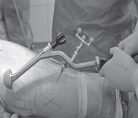
The minimally invasive Micro-hip® approach it derived from the Smith-Petersen-procedure. Landmarks for orientation are the greater trochanter, anterior superior iliac spine and the iliotibial tract. The incision is running in the ventral part of the iliotibial tract with his distal point in the middle of the great trochanter, running 6–8 cm proximally at the ventral rim of the iliotibial tract. When incising the subcutis the ventral rim of the iliotibial tract can be palpated with its stronger tissue compare to the muscle fascia. The ilitobial tract is incised, the tensor fibers and the gluteal muscle are digitally mobilized to reach the interval with the rectus muscle. Smith-Petersen described this approach using of the anterior iliofemoral section. This way you reach the joint capsule without splitting muscles or tendons. The hip joint is not dislocated. The osteotomy of the femoral neck is performed in situ. After removing the head proximal femur and the acetabulum can be exposed easily. For this procedure only standard instruments are used. After removing the acetabular labrum and releasing contractures of the capsule the acetabular reamer is inserted. This is a special instrument with angulation. With good sight to the deep structures we impact a press-fit acetabular component and a cement-free stem, or, if necessary, a cemented stem.
/16/16.jpg)
In order to check stability the leg can be moved in all directions: flexion, extension, abduction, rotation. In all our hip-replacements we perform an intraoperative X-ray-control, which allows additionally checking size and position of the implant and leg length.
Outcome Measurement
The duration of the operation lasts from 45 to 90 minutes (77 minutes). Blood loss, during the first 24 hours, including the intraoperative bleeding, sums up to 300 ml in average.
Patients are mobilized already on the first day after operation with two crutches. Due to cement-free-implants we ask our patients to use their crutches for 5 weeks postoperatively.
Radiographic evaluation is done in a standard anteroposterior pelvic and a lateral hip position. Harris Hip Score increased from 68.5 preoperatively to 92 after one year postoperatively.
Cups are implanted in an inclination of 47° (± 5.2°) and about 10 % had a stem varus/valgus of more than 3° [1].
We don’t see any specific complication, except single patients having lateral femoral cutaneous nerve palsy due to the incision, which is normally not realized by the patient, but is detected by exact clinical examination.
/16/16_2.jpg)
Discussion and conclusion our measurement show a reduced blood loss with this operative technique and the possibility to mobilize the patient early postoperatively nearly without any pain due to the completely kept muscles. This gains improved functional results already in the early postoperative period. We can underline the statements, that the minimized muscle trauma is beneficial for recovery and function [2]. It is important to point out, that our operative technique is not to be compared to the mini-hip in Watson-Jones technique [3]. We also like to underline, that our technique is not comparable to these procedures, which go through the tensor fascia latae muscle [4].
Although our technique has a difficult learning curve, our results reveal the advantages of the intraoperative procedure and the quick postoperative recovery of the patient.
1. Sendtner E., Borowiak K., Schuster T., Woerner M., Grifka J., Renkawitz T. Tackling the learning curve: comparison between the anterior, minimally invasive (Micro-hip®) and the lateral, transgluteal (Bauer) approach for primary total hip replacement // Arch. Orthop. Trauma Surg. — 2010. — 131. — 597-602.
2. Leuchte S., Riedl K., Wohlrab D. Immediate post-operative advantages of minimally invasive hip replacement-results of symmetry and load from the measurement of ground reaction force // Z. Orthop. Unfall. — 2009. — 147(1). — 69-78.
3. Bertin K.C., Rottinger H. Anterolateral mini-incision hip replacement surgery: a modified Watson-Jones approach // Clin. Orthop. Relat. Res. — 2004. — 429. — 248-255.
4. Meneghini R.M., Pagnano M.W., Trousdale R.T., Hozack W.J. Muscle damage during MIS total hip arthroplasty: Smith-Petersen versus posterior approach // Clin. Orthop. Relat. Res. — 2006. — 453. — 293-298.